Product Sheet CP10246
Description
BACKGROUND The Wnt genes encode a large, highly conserved family of cysteine-rich secreted glycoproteins that play essential roles in controlling tissue patterning, cell fate and proliferation from Drosophila to humans. There are 19 mammalian Wnt proteins identified and all bind to frizzled (Fzd) cell surface receptors to initiate cellular responses. The members exhibit unique expression patterns and distinct functions in development. They can be divided into three distinct types based on their ability to induce transformation of the mouse mammary epithelial cell line C57MG. The highly transforming members include Wnt-1, Wnt-3a, and Wnt-7a. The intermediately transforming members include Wnt-2, Wnt-5b, and Wnt-7b and nontransforming members are Wnt-4, Wnt-5a, Wnt-6, and Wnt-11. They signal through either the canonical pathway or at least two different noncanonical pathways. In short, the canonical Wnts signal through nuclear beta-catenin/Tcf-Lef while the noncanonical Wnts function through alternate signaling cascades that include Ca++/PKC, RhoA/JNK or RhoB/Rab4. In addition to secreted Wnt glycoproteins, the Wnt signaling network also includes several extracellular secreted regulators that antagonize Wnt actions including secreted frizzledrelated proteins (Sfrps), Wnt inhibitory factors (Wif), and the dickkopf (Dkk) proteins which specifically block canonical Wnt signaling. Mutations in Wnt genes or Wnt pathway components lead to specific developmental defects, while various human diseases, including cancer, are caused by abnormal Wnt signaling.1
Wnt5a is a noncanonical Wnt ligand involved in outgrowth of multiple structures during vertebrate development. It activates the noncanonical Wnt signaling pathways involved in PKC and CamKII as well as PKD and JNK. However in some contexts, Wnt5a can also act similar to other Wnt family members and bind with Frizzled (Fz) receptors located on the cell surface and activates cytosolic Dishevelled (Dsh) proteins, leading to stabilization of beta-catenin. Accumulation of beta-catenin results in its nuclear translocation and activation of Lef /Tcf transcription factors. Additionally, studies showed that Wnt-5a signaling can antagonize the canonical Wnt signaling pathway by promoting beta-catenin degradation. Moreover, the beta-catenin degradation induced by Wnt-5a is independent of phosphorylation by GSK-3 and involves the induction of Siah2 expression. And Wnt-5a–induced beta-catenin degradation does not require activation of CaMKII or NF-AT.2
Targeted deletion of Wnt5a−/− in mice resulted in perinatal lethality demonstrating a critical role for normal development of structures necessary for life. It was demonstrated that Wnt5a plays an essential role in outgrowth and patterning along the proximal–distal axis. Furthermore, Wnt5a has been shown to have growth-enhancing or oncogenic potential: Wnt5a upregulates proliferation of progenitor cells common to many structures in mice, Wnta5a expression is upregulated in a variety of human primary tumor samples, and WNT5A facilitates cell invasion in human metastatic melanoma. However, it was also shown that Wnt5a negatively regulates B cell proliferation in a cell-autonomous manner. Deletion of the Wnt5a gene, loss of Wnt5a expression, and increased cyclin D1 expression is observed in mouse and human B cell lymphomas and myeloid leukemias. Wnt5A can function as a tumor suppressor.3 In addition, it was discovered that post-synaptic damage induced by Abeta oligomers in hippocampal neurons is prevented by non-canonical Wnt5A pathway activation.4
REFERENCES
1. Wodarz, A. et al: Annu. Rev. Cell Dev. Biol. 14: 59–88, 1998
2. Topol, L. et al: J. Cell Biol.162:899-908, 2003
3. Liang, H. et al: Cancer Cell 4:349-60, 2003
4. Cerpa, W. et al: Mol. Neurodegenr. 5:3, 2010
2. Topol, L. et al: J. Cell Biol.162:899-908, 2003
3. Liang, H. et al: Cancer Cell 4:349-60, 2003
4. Cerpa, W. et al: Mol. Neurodegenr. 5:3, 2010
Products are for research use only. They are not intended for human, animal, or diagnostic applications.
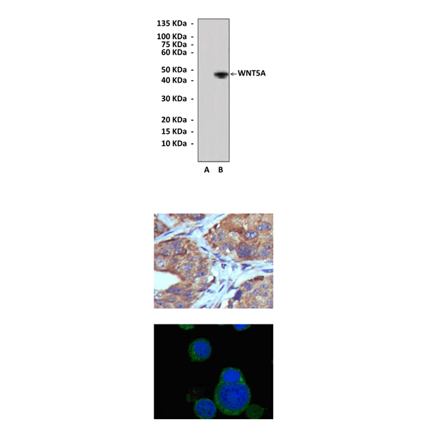
Top: Western Blot detection of WNT5A proteins in 293 cell lysates transfected with human WNT5A expression construct or mock vector using WNT5A Antibody. Middle: This antibody stains paraffin-embedded human lung cancer tissue in immunohistochemical analysis. Bottom: It also stains PC-12 cells in confocal immunofluorescent testing (WNT5A Antibody: Green; DRAQ5 DNA dye: Blue).
Details
Cat.No.: | CP10246 |
Antigen: | Purified recombinant human WNT5A fragments expressed in 293 cells. |
Isotype: | Mouse IgG1 |
Species & predicted species cross- reactivity ( ): | Human, Mouse, Rat |
Applications & Suggested starting dilutions:* | WB 1:1000 IP n/d IHC 1:200 ICC 1:200 FACS n/d |
Predicted Molecular Weight of protein: | 45 kDa |
Specificity/Sensitivity: | Detects endogenous WNT5A proteins without cross-reactivity with other family members. |
Storage: | Store at -20°C, 4°C for frequent use. Avoid repeated freeze-thaw cycles. |
*Optimal working dilutions must be determined by end user.
Products
| Product | Size | CAT.# | Price | Quantity |
|---|---|---|---|---|
| Rabbit WNT5A Antibody: Rabbit WNT5A Antibody | Size: 100 ul | CAT.#: CP10246 | Price: $413.00 |