MSDS Cryopreserved Cells
Instructions HEM
Cell Apps Flyer Skin Cells
5 Important Cell Culture Rules
Cell Apps Poster Primary Cells
Cell Applications Inc Brochure

Description

HEM
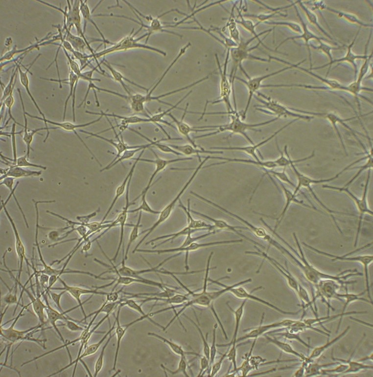
Human Epidermal Melanocytes (HEM) from Cell Applications, Inc. maintain their characteristic shape in culture for many generations.  They produce melanin and serve as a useful cell model for the studies of melanocyte proliferation and differentiation, as well as progression of melanocytic neoplasia. Epidermal melanocytes are pigment-producing cells located at the basal level of epidermis, where they interact with keratinocytes via cellular processes called dendrites.  Melanin, the pigment produced by melanocytes and responsible for skin color, is then transferred to keratinocytes, where it is stored in vesicles called melanosomes located around the nucleus to provide protection from UV radiation.
HEM provided by Cell Applications, Inc. have been used to:
  • Identify unique features and biomarkers of melanoma cells
  • Show that IGFBP7 is dispensable for B-RAFV600E-induced senescence
  • Investigate mechanisms of cellular senescence, in particular by showing that Id1 extends the life span of melanocytes through inhibition of p16/INK4a expression
  • Discover the central role of oncogenic BRAF gene in melanoma oncogenesis by demonstrating that the constitutive MAPK pathway activation leading to activation of mTOR, STAT3-dependent transcription of Mcl-1, Tbx3-mediated repression of E-cadherin leading to increased metastasis in melanoma cells all result from overexpression and/or mutations of BRAF gene
  • Discover other key players in oncogenic signaling leading to melanoma, such as PI3K which regulates MAPK activation in response to oncogenic c-Kit activity, Nck2 adaptor protein which participates in  regulation of tyrosine kinases activity and the role of mitochondria metabolism in advanced melanoma
  • Demonstrate that p16INK4a-Rb-CDK4/6 senescence-inducing tumor suppressor pathway in inhibited in melanoma cells leading to proliferation of cells harboring DNA damage and by studying expression of senescence markers
(Click to Enlarge) Human Epidermal Melanocytes: HEM.  Pigment producting cells isolated from skin tissue.

Details

Tissue
Normal healthy human neonatal foreskin
QC
No bacteria, yeast, fungi, mycoplasma, virus
Bioassay
Attach, spread, proliferate in Growth Med
Cryovial
500,000 HEM (2nd passage) frozen in Basal Medium w/ 30% FBS, 10% DMSO
Kit
Cryovial frozen HEM (104-05n), Growth Medium (135-500), Subcltr Rgnt Kit (090K)
Proliferating
Shipped in Gr Med, 3rd psg (flasks or plates) 
Doublings
At least 12
Applications
Laboratory research use only (RUO). Not for human, clinical, diagnostic or veterinary use.
Instructions HEM

Format: PDF

Download Now
MSDS Cryopreserved Cells

Format: PDF

Download Now

Products

Product Size CAT.# Price Quantity
Cryopreserved Cryopreserved HEM, NeonatalHuman Epidermal Melanocytes Total Kit, neonatal: 5x10^5 Cells (Neonatal), Medium & Subculture Reagents (See Details tab for specifics) Size: 1 Kit CAT.#: 104K-05n Price: $698.00
-+
Cryopreserved Epidermal Melanocyte (HEM), neonatal: Frozen HEM (5x10^5) Size: 1 Cryovial CAT.#: 104-05n Price: $525.00
-+
Proliferating Epidermal Melanocyte (HEM), neonatal: Actively growing, dividing cells, in medium Size: T-25 Flask CAT.#: 105-25n Price: $525.00
-+
Proliferating Epidermal Melanocyte (HEM), neonatal: Actively growing, dividing cells, in medium Size: T-75 Flask CAT.#: 105-75n Price: $715.00
-+
Proliferating Epidermal Melanocyte (HEM), neonatal: Actively growing, dividing cells, in medium Size: 24 Well CAT.#: 105-24Wn Price: $715.00
-+
Proliferating Epidermal Melanocyte (HEM), neonatal: Actively growing, dividing cells, in medium Size: 96 Well CAT.#: 105-96Wn Price: $835.00
-+
Cryopreserved Human Epidermal Melanocytes Total Kit, adult: 5x10^5 Cells (Adult), Medium & Subculture Reagents (See Details tab for specifics) Size: 1 Kit CAT.#: 104K-05a Price: $718.00
-+
Cryopreserved Epidermal Melanocyte (HEM), adult: Frozen HEM (5x10^5) Size: 1 Cryovial CAT.#: 104-05a Price: $545.00
-+
Proliferating Epidermal Melanocyte (HEM), adult: Actively growing, dividing cells, in medium Size: T-25 Flask CAT.#: 105-25a Price: $545.00
-+
Proliferating Epidermal Melanocyte (HEM), adult: Actively growing, dividing cells, in medium Size: T-75 Flask CAT.#: 105-75a Price: $735.00
-+
Proliferating Epidermal Melanocyte (HEM), adult: Actively growing, dividing cells, in medium Size: 24 Well CAT.#: 105-24Wa Price: $735.00
-+
Proliferating Epidermal Melanocyte (HEM), adult: Actively growing, dividing cells, in medium Size: 96 Well CAT.#: 105-96Wa Price: $855.00
-+

Related Products

Product Size CAT.# Price Quantity
HEM Growth Medium: All-in-one ready-to-use Size: 500 ml CAT.#: 135-500 Price: $120.00
-+
HEM Growth Medium Kit: Basal medium & growth supplement sold together packaged separately Size: Yields 500 ml CAT.#: 135K-500 Price: $130.00
-+
HEM Basal Medium: Basal medium (contains no growth supplement).  Add GS before use. Size: 500 ml CAT.#: 134-500 Price: $74.00
-+
HEM Growth Supplement: Added to Basal Medium to create Growth Medium Size: 5 ml (3 parts) CAT.#: 135-GS Price: $74.00
-+
Freezing Medium for HEM: Freezing Medium optimized for the unique needs of Human Epidermal Melanocytes (HEM). Size: 50 ml CAT.#: 040-50HEM Price: $75.00
-+

Extended Family Products

Product Size CAT.# Price Quantity
Human Epidermal Growth Factor (EGF): Human Epidermal Growth Factor Size: 100 ug CAT.#: RP1026-100 Price: $86.00
-+
Human Epidermal Growth Factor (EGF): Human Epidermal Growth Factor Size: 500 ug CAT.#: RP1026-500 Price: $194.00
-+
Human Epidermal Growth Factor (EGF): Human Epidermal Growth Factor Size: 1000 ug CAT.#: RP1026-1000 Price: $264.00
-+
Epidermal Melanocyte RNA (HEM RNA), Neonatal: Total RNA prepared from Human Epidermal Melanocytes, neonatal Size: 10 ug CAT.#: 104-R10n Price: $398.00
-+
Epidermal Melanocyte RNA (HEM RNA), Neonatal: Total RNA prepared from Human Epidermal Melanocytes, neonatal Size: 25 ug CAT.#: 104-R25n Price: $796.00
-+
Subculture Reagent Kit: 100 ml each of HBSS, Trypsin/EDTA & Trypsin Neutralizing Solution Size: 1 Kit CAT.#: 090K Price: $63.00
-+
Human EGF, Animal-Free: Human Epidermal Growth Factor, Animal-Free Size: 100 ug CAT.#: RP1026AF-100 Price: $95.00
-+
Human EGF, Animal-Free: Human Epidermal Growth Factor, Animal-Free Size: 500 ug CAT.#: RP1026AF-500 Price: $213.00
-+
Human EGF, Animal-Free: Human Epidermal Growth Factor, Animal-Free Size: 1000 ug CAT.#: RP1026AF-1000 Price: $290.00
-+

Resources/Documents

Cell Apps Flyer Skin Cells

Format: PDF

Downoad Now
5 Important Cell Culture Rules

Format: PDF

Downoad Now
Cell Apps Poster Primary Cells

Format: PDF

Downoad Now
Cell Applications Inc Brochure

Format: PDF

Downoad Now

Publications

2015
Wiersma, V., M. Bruyn, C. Shi, M. Gooden, M. Wouters, D. Samplonius, D. Hendriks, H. Nijman, Y. Wei, J. Zhou, W. Helfrich, and E. Bremer. 2015. C-type lectin-like molecule-1 (CLL1)-targeted TRAIL augments the tumoricidal activity of granulocytes and potentiates therapeutic antibody-dependent cell-mediated cytotoxicity. mAbs, Vol 7, 11 March.
2014
Todd, J., L. Scurr, T. Becker, R. Kefford and H. Rizos. 2014. The MAPK pathway functions as a redundant survival signal that reinforces the PI3K cascade in c-Kit mutant melanoma. Oncogene, 33:236-245. HEM
2013
Becker, T.M., S.C. Boyd, B. Mijatov, K. Gowrishankar, S. Snoyman, G.M. Pupo, R.A. Scolyer, G.J. Mann, R.F. Kefford, X.D. Zhang, and H. Rizos. 2013. Mutant B-RAF-Mcl-1 survival signaling depends on the STAT3 transcription factor. Oncogene. doi: 10.1038/onc.2013.45.
Boyd, S.C., B. Mijatov, G.M. Pupo, S.L. Tran, K. Gowrishankar, H.M. Shaw, C.R. Goding, R.A. Scolyer, G.J. Mann, R.F. Kefford, H. Rizos, and T.M. Becker. 2013. Oncogenic B-RAFV600E Signaling Induces the T-Box3 Transcriptional Repressor to Repress E-Cadherin and Enhance Melanoma Cell Invasion. J Invest Dermatol. 133:1269-1277.
Fung, C., G.M. Pupo, R.A. Scolyer, R.F. Kefford, and H. Rizos. 2013. p16INK4a deficiency promotes DNA hyperreplication and genetic instability in melanocytes. Pigment cell & melanoma research. 26:236-246.
Romeo, Y., J. Moreau, P.J. Zindy, M. Saba-El-Leil, G. Lavoie, F. Dandachi, M. Baptissart, K.L.B. Borden, S. Meloche, and P.P. Roux. 2013. RSK regulates activated BRAF signalling to mTORC1 and promotes melanoma growth. Oncogene. 32:2917-2926.
2012
Barbi de Moura, M., G. Vincent, S.L. Fayewicz, N.W. Bateman, B.L. Hood, M. Sun, J. Suhan, S. Duensing, Y. Yin, C. Sander, J.M. Kirkwood, D. Becker, T.P. Conrads, B. Van Houten, and S.J. Moschos. 2012. Mitochondrial Respiration - An Important Therapeutic Target in Melanoma. PloS one. 7:e40690.
De Moura, M., G. Vincent, S. Fayewicz, N. Bateman, B. Hood, M. Sun, J. Suhan, S. Duensing, Y. Yin, C. Sander, J. Kirkwood, D. Becker, T. Conrads, B. Van Houten and S. Moschos. 2012. Mitochondrial Respiration - An Important Therapeutic Target in Melanoma. PLoS ONE, dx.doi.org/10.1371/journal.pone.0040690.
Ho, J., M. de Moura, Y. Lin, G. Vincent, S. Thorne, L. Duncan, L. Hui-Min, J. Kirkwood, D. Becker, B. Van Houten and S. Moschos. 2012. Importance of glycolysis and oxidative phosphorylation in advanced melanoma.  Molec Cancer, 11:76.
Todd, J., L. Scurr, T. Becker, R. Kefford, and H. Rizos. 2012. The MAPK pathway functions as a redundant survival signal that reinforces the PI3K cascade in c-Kit mutant melanoma. Oncogene. doi: 10.1038/onc.2012.1562.
Tran, S.L., S. Haferkamp, L.L. Scurr, K. Gowrishankar, T.M. Becker, C. Desilva, J.F. Thompson, R.A. Scolyer, R.F. Kefford, and H. Rizos. 2012. Absence of distinguishing senescence traits in human melanocytic nevi. J. Inv. Dermatol. 132:2226-2234.
Villareal, M.O., J. Han, K. Ikuta, and H. Isoda. 2012. Mechanism of Mitf inhibition and morphological differentiation effects of hirsein A on B16 melanoma cells revealed by DNA microarray. Journal of dermatological science. 67:26-36.
Yajima, I., M.Y. Kumasaka, Y. Naito, T. Yoshikawa, H. Takahashi, Y. Funasaka, T. Suzuki, and M. Kato. 2012. Reduced GNG2 expression levels in mouse malignant melanomas and human melanoma cell lines. American J. Cancer Res. 2:322.
2011
Labelle-Côté, M., J. Dusseault, S. Ismaïl, A. Picard-Cloutier, P. Siegel, and L. Larose. 2011. Nck2 promotes human melanoma cell proliferation, migration and invasion in vitro and primary melanoma-derived tumor growth in vivo. BMC cancer. 11:443.
Weber, D., J. Markowitz, F. Carrier, and A. Mackerell. 2011. Inhibitors of the S100-p53 protein-protein interaction and method of inhibiting cancer employing the same. Patent US 8053477 B2.  
2010
Mahmoud, M., A. Hesham, Y. Ahmed, and M. Sayed. 2010. Inhibition of melanogenesis by the extract from Agaricus blazei without affecting iNOS gene expression. World J Microbiol Biotechnol, DOI 10.1007/s11274-010-0387-6.
Ohshima, Y., I Yajima, K. Takeda, M. Lida, M. Kumasaka, Y. Matsumoto, and M. Kato. 2010. c-RET molecule in malignant melanoma from oncogenic RET-carrying transgenic mice and human cell lines. PLoS One 5: e10279.
Scurr, L.L., G.M. Pupo, T.M. Becker, K. Lai, D. Schrama, S. Haferkamp, M. Irvine, R.A. Scolyer, G.J. Mann, and J.C. Becker. 2010. IGFBP7 is not required for B-RAF-induced melanocyte senescence. Cell. 141:717-727.
2008
Silveira, J., M. Pereda, S. Eberlin, G. Dieamant, and L. Di Stasi. 2008. Effects of Coccoloba uvifera L. on UV-stimulated melanocytes. Photodermatology, Photoimmunology & Photomedicine, 24:308-313.
2006
Baron, U., I. Turbachova, A. Hellwag, F. Eckardt, K. Berlin, U. Hoffmüller, P. Gardina, and S. Olek. 2006.  DNA Methylation Analysis as a Tool for Cell Typing. Epigenetics 1:55-60.
2005
Weber, D., J. Markowitz, F. Carrier, and A. MacKerell. 20015. Inhibitors of the S100-p53 protein-protein interaction and method of inhibiting cancer employing the same. Patent Application US 20060004085 A1.
2004
Tanami, H., I. Imoto, A. Hirasawa, Y. Yuki, I. Sonoda, J. Inoue, K. Yasui, A. Misawa-Furihata, Y. Kawakami, and J. Inazawa. 2004. Involvement of overexpressed wild-type BRAF in the growth of malignant melanoma cell lines. Oncogene, 23:8796-8804.