Product Sheet CP10416
Description
BACKGROUND NF-kappa-B is a pleiotropic transcription factor which is present in almost all cell types and is involved in many biological processed such as inflammation, immunity, differentiation, cell growth, tumorigenesis and apoptosis. The NF-kappa-B family includes five members, p65 (Rel-A), c-Rel, Rel-B, NF-kappa-B1 (p50 and its precursor p105), and NF-kappa-B2 (p52 and its precursor p100). In the inactive state, NF-kappa-B proteins are sequestered in the cytoplasm by I B proteins (IkappaBalpha, IkappaBbeta, IkappaBepsilon, and IkappaBgamma). IkappaB inactivates NF-kappaB by masking the nuclear localization signals (NLS). Following stimulation, I B kinase (IKK) complexes are activated to phosphorylate IkappaB proteins, which leads to proteasome-mediated degradation of IkappaB proteins. The released NF-kappa-B proteins translocate into the nucleus where they bind to kappaB sequences in the promoters of target genes to initiate transcription.1 In general, activated NF-kappa-B dimers containing p65, c-Rel, or Rel-B can transactivate NF-kappa-B -dependent genes. In contrast, NF-kappa-B homodimers, p50/p50 and p52/p52, which lack transactivation domains, function primarily to inhibit NF-kappa-B -responsive genes. However, binding of p50/p50 or p52/p52 homodimers to B cell lymphoma 3 (Bcl-3), a transcriptional coactivator, confers the ability of these homodimers to induce NF-kappa-B responsive genes. Bcl-3 belongs to the IkappaB family and can interact with NF-kappa-B proteins through its ankyrin repeats. Unlike other IkappaB proteins, which are expressed in the cytoplasm and function as repressors of NF-kappa-B, Bcl-3 is predominately expressed in the nucleus and functions as an activator through interactions with p50 and p52 homodimers.2 NF-kaapa-B1 appears to have dual functions such as cytoplasmic retention of attached NF-kappa-B proteins by p105 and generation of p50 by a cotranslational processing. The proteasome-mediated process ensures the production of both p50 and p105 and preserves their independent function, although processing of NF-kappa-B1/p105 also appears to occur post-translationally. p50 binds to the kappa-B consensus sequence 5\'-GGRNNYYCC-3\', located in the enhancer region of genes involved in immune response and acute phase reactions.3
In addition to regulation of NF-kappa-B activity through removal of IkappaB from NF-kappa-B/IkappaB complexes, NF-kappa-B activity is also regulated through modulation of its transcriptional function. Changes in NF-kappa-B transcriptional activity have been assigned to inducible phosphorylation of the p65 subunit at Ser276, Ser529, and Ser536 by a large variety of kinases in response to different stimuli.4 Additionally, NF-kappa-B -dependent transcription requires multiple coactivators possessing histone acetyltransferase activity: CREB binding protein (CBP) and its homolog p300, p300/CBP-associated factor (P/CAF), SRC-1/NcoA-1, and TIF-2/GRIP-1/NcoA-2. Importantly, recruitment of CBP is enhanced by phosphorylation by the catalytic subunit of PKA (PKAc) of p65 at Ser276. More recently, other findings demonstrated a role for histone deacetylases (HDACs) as well. The first evidence came from the demonstration that inhibition of HDAC activity by trichostatin A (TSA) increases NF-kappa-B-dependent gene expression. It was next shown that NF-kappa-B interacts with distinct HDAC isoforms to negatively regulate gene expression, presumably through the deacetylation of histones and/or nonhistone proteins. Importantly, the phosphorylation status of p65 determines whether it associates with CBP/p300 or HDAC-1, ensuring that only signal-induced NF-kappa-B entering the nucleus can activate transcription.5
In addition to regulation of NF-kappa-B activity through removal of IkappaB from NF-kappa-B/IkappaB complexes, NF-kappa-B activity is also regulated through modulation of its transcriptional function. Changes in NF-kappa-B transcriptional activity have been assigned to inducible phosphorylation of the p65 subunit at Ser276, Ser529, and Ser536 by a large variety of kinases in response to different stimuli.4 Additionally, NF-kappa-B -dependent transcription requires multiple coactivators possessing histone acetyltransferase activity: CREB binding protein (CBP) and its homolog p300, p300/CBP-associated factor (P/CAF), SRC-1/NcoA-1, and TIF-2/GRIP-1/NcoA-2. Importantly, recruitment of CBP is enhanced by phosphorylation by the catalytic subunit of PKA (PKAc) of p65 at Ser276. More recently, other findings demonstrated a role for histone deacetylases (HDACs) as well. The first evidence came from the demonstration that inhibition of HDAC activity by trichostatin A (TSA) increases NF-kappa-B-dependent gene expression. It was next shown that NF-kappa-B interacts with distinct HDAC isoforms to negatively regulate gene expression, presumably through the deacetylation of histones and/or nonhistone proteins. Importantly, the phosphorylation status of p65 determines whether it associates with CBP/p300 or HDAC-1, ensuring that only signal-induced NF-kappa-B entering the nucleus can activate transcription.5
REFERENCES
1. Hayden, M.S. & Ghosh, S.: Cell 134:344-62, 2008
2. Dai, R. et al: J. Immunol.179:1776-83, 2007
3. Moorthy, A.K. et al: EMBO J. 25:1945-56, 2006
4. Vermeulen, L. et al: Biochem. Pharmacol. 64:673-90, 2002
5. Zhong, H. et al: Mol. Cell 9:625-36, 2002
2. Dai, R. et al: J. Immunol.179:1776-83, 2007
3. Moorthy, A.K. et al: EMBO J. 25:1945-56, 2006
4. Vermeulen, L. et al: Biochem. Pharmacol. 64:673-90, 2002
5. Zhong, H. et al: Mol. Cell 9:625-36, 2002
Products are for research use only. They are not intended for human, animal, or diagnostic applications.
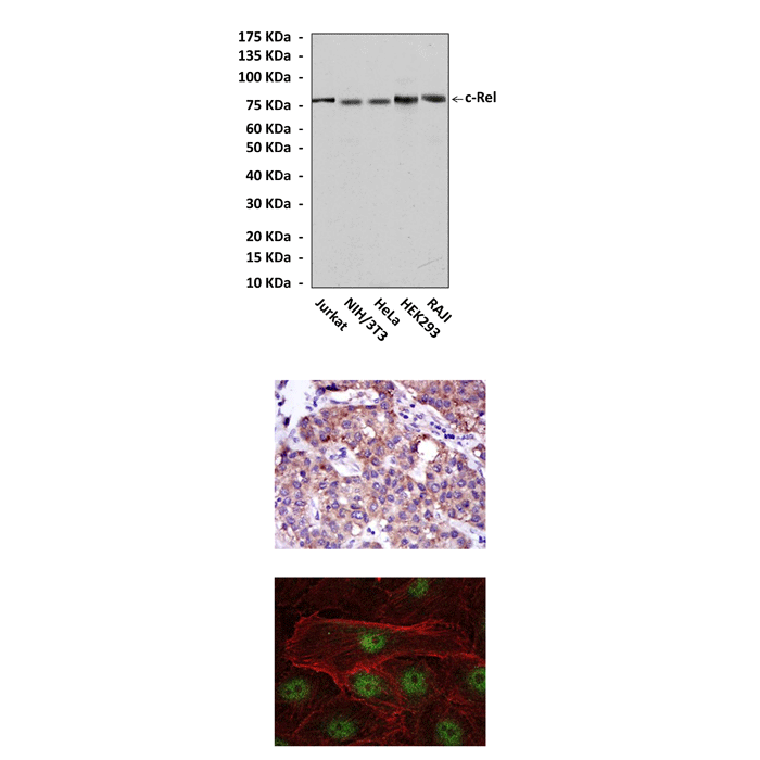
(Click to Enlarge) Top: Western blot detection of c-Rel proteins in various cell lysates using c-Rel Antibody. Middle: This antibody stains paraffin-embedded human liver cancer tissue in IHC analysis. Bottom: It also stains U251 cells in confocal immunofluorescent testing (c-Rel Antibody: Green; Actin filaments: Red).
Details
Cat.No.: | CP10416 |
Antigen: | Raised against recombinant human c-Rel fragments expressed in E. coli. |
Isotype: | Mouse IgG1 |
Species & predicted species cross- reactivity ( ): | Human, Mouse, Rat |
Applications & Suggested starting dilutions:* | WB 1:1000 IP 1:25 - 1:50 IHC 1:50 - 1:200 ICC 1:50 - 1:200 FACS n/d |
Predicted Molecular Weight of protein: | 78 kDa |
Specificity/Sensitivity: | Detects c-Rel proteins in various cell lysate. |
Storage: | Store at -20°C, 4°C for frequent use. Avoid repeated freeze-thaw cycles. |
*Optimal working dilutions must be determined by end user.
Products
| Product | Size | CAT.# | Price | Quantity |
|---|---|---|---|---|
| Mouse c-Rel Antibody: Mouse c-Rel Antibody | Size: 100 ul | CAT.#: CP10416 | Price: $457.00 |