Description
BACKGROUND ICAM-1(intercellular adhesion molecule-1) is a transmembrane glycoprotein of 532 amino acids, which has a molecular mass ranging from 90 to 110 kDa. ICAM-1 is a member of the immunoglobulin supergenge family, and contains five extracellular immunoglobulin-like domains. It is expressed on various cells of both hematopoietic and non-hematopoietic cells, including endothelial cells, leukocytes other than basophilic granulocytes, T cells, B cells, fibroblasts, and cancer cells. As a member of adhesion molecules, ICAM-1 binds to two integrins belonging to the beta2 subfamily CD11a/CD18 (LFA-1) and CD11b/CD18 (Mac-1) on the surface of leukocytes. ICAM1 is a key molecule in immune-mediated and inflammatory processes and function as a co-stimulatory signal which is important for the trans-endothelial migration of leukocytes and the activation of T cells.1 During leukocyte trans-endothelial migration, ICAM1 engagement promotes the assembly of endothelial apical cups through SGEF and RHOG activation. Moreover, ICAM-1 has a crucial role in the induction of an immune response and is instrumental in migration of T cells into inflamed tissue. ICAM-1 on target cells leads to recruitment of the MHC-I proteins to the contact area and enhances presentation of cognate peptide MHC-I complex to cytotoxic T cells. ICAM-1 interacts with important factors involved in many kinds of human cancers. ICAM-1 is involved in transmembrane signal transduction in the regulatory process of cell proliferation through the mitogen-activated protein kinase (MAPK) pathway and eventually the AP-1 pathway.2 ICAM-1 can be induced under inflammatory condition by TNF-alpha in a process that involves IKK-beta.
In addition, inhibition of ICAM-1 expression on melanoma cells reduces the metastatic ability of the melanoma cells, indicating an important role of ICAM-1 in metastasis.3 The existence of a soluble variant of ICAM-1 in the circulation has been described, with elevated levels being reported in several diseases; higher levels being associated with liver metastases in gastric, colonic, gall bladder and pancreatic cancer, and with reduced survival in patients with malignant melanoma. In case of rhinovirus infection, ICAM-1 acts as a cellular receptor for the virus.4
In addition, inhibition of ICAM-1 expression on melanoma cells reduces the metastatic ability of the melanoma cells, indicating an important role of ICAM-1 in metastasis.3 The existence of a soluble variant of ICAM-1 in the circulation has been described, with elevated levels being reported in several diseases; higher levels being associated with liver metastases in gastric, colonic, gall bladder and pancreatic cancer, and with reduced survival in patients with malignant melanoma. In case of rhinovirus infection, ICAM-1 acts as a cellular receptor for the virus.4
REFERENCES
1. Van Seventer, G.A. et al: J. Immunol. 144:4579-86, 1990
2. Chen, C.C. et al: Cell Signal. 13:543-53, 2001
3. Miele, M.E. et al: Exp Cell Res. 214:231-41, 1994
4. Greve, J.M. et al: Cell 56:839-47, 1989
2. Chen, C.C. et al: Cell Signal. 13:543-53, 2001
3. Miele, M.E. et al: Exp Cell Res. 214:231-41, 1994
4. Greve, J.M. et al: Cell 56:839-47, 1989
Products are for research use only. They are not intended for human, animal, or diagnostic applications.
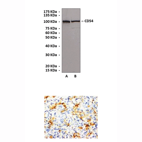
Top: Detection of ICAM-1 proteins from HUVEC cells (A)(201p-25n) and Human Lymphatic endothelial cell (B)(100LK-25a) lysates in Western blot assay using Anti-ICAM-1 Antibody. Bottom: Immunohistochemical staining of paraffin-embedded rat spleen tissue, using Anti-ICAM-1.
Details
Cat.No.: | CA0541 |
Antigen: | Peptide mapping at the C-terminal region of the human ICAM-1. This antibody was purified by peptide affinity chromatography. |
Isotype: | Affinity-purifiedrabbit polyclonal IgG |
Species & predicted species cross- reactivity ( ): | Human, Rabbit, Rat, Mouse |
Applications & Suggested starting dilutions: | WB 1:500 - 1:1000 IP n/d IHC (Paraffin) 1:50 - 1:200 ICC n/d FACS n/d |
Predicted Molecular Weight of protein: | 90-110 kDa |
Specificity/Sensitivity: | Reacts specifically with ICAM-1 and does not cross react with other members of the ICAM family. |
Storage: | Store at 4° C for frequent use; at -20° C for at least one year. |
*Optimal working dilutions must be determined by end user.
Products
| Product | Size | CAT.# | Price | Quantity |
|---|---|---|---|---|
| Anti-ICAM-1: Rabbit Intercellular Adhesion Molecule-1 Antibody | Size: 100 ul | CAT.#: CG1238 | Price: $275.00 |
Publications
2014
Trauger, R. 2014. Felinized Antibodies and Methods of Treating Retroviral Infections in Felines. Patent Application US 20140348829 A1.
2012
Trauger, R. 2012. Methods of Treating Retroviral Infections in Felines. Patent Application US 20140140997 A1.