Product Sheet CC1011
Description
BACKGROUND Eph family of receptor tyrosine kinases consists of at least 14 distinct receptors and has eight membrane-bound ligands, known as the ephrins. This is the largest family of receptor tyrosine kinases. Eph proteins are divided into two subfamilies: the EphA receptors (A1-A8) that bind glycosyl phosphatidylinositol (GPI)-linked ephrin-A ligands (A1-A5), and the EphB receptors (B1-B6) that bind transmembrane ephrin-B ligands (B1-B3). The only known crosstalk between the A and B subfamilies occurs with the EphA4 receptor, which can bind ephrins-B2 and -B3 as well as the entire A subclass. There is a great deal of redundancy of receptor-ligand binding specificity within each subfamily, although binding affinities vary.1,2 One of the unique features of Eph/ephrin signaling is the fact that both receptors and ligands are competent to transduce a signaling cascade upon interaction. Eph-activated signaling is termed forward, and ephrin-activated signaling is termed reverse. Another level of complexity stems from the fact that interactions between Eph receptors and ephrins can happen in trans (between two opposing cells) or in cis (within the same cell). It is commonly assumed that trans interactions are activating while cis interactions are inhibiting.3 EphA2 is a transmembrane receptor tyrosine kinase that is up-regulated on many aggressive carcinoma cells. Despite its overexpression, the EphA2 on malignant cells fails to bind its ligand, ephrinAl, which is anchored to the membrane of adjacent cells. Unlike other receptor kinases, EphA2 demonstrates kinase activity that is independent of ligand binding. However, ligand binding causes EphA2 to negatively regulate tumor cell growth and migration.4
REFERENCES
1. Brantley-Sieders, D.M. & Chen, J.: Angiogenesis. 7:17, 2004.
2. Murai, K.K. & Pasquale, E.B.: J. Cell Sci. 116:2823, 2003.
3. Arvanitis, D. & Davy, A.: Genes & Dev. 22:416, 2008.
4. Carles-Kinch, K. et al.: Cancer Res. 62:2840, 2002.
1. Brantley-Sieders, D.M. & Chen, J.: Angiogenesis. 7:17, 2004.
2. Murai, K.K. & Pasquale, E.B.: J. Cell Sci. 116:2823, 2003.
3. Arvanitis, D. & Davy, A.: Genes & Dev. 22:416, 2008.
4. Carles-Kinch, K. et al.: Cancer Res. 62:2840, 2002.
Products are for research use only. They are not intended for human, animal, or diagnostic applications.
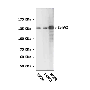
(Click to Enlarge) Various cell lysates were subjected to Western Blot analysis using EphA2 Antibody.
Details
Cat.No.: | CC1011 |
Antigen: | An epitope near the human EphA2 carboxyl terminal sequence. |
Isotype: | Rabbit polyclonal IgG |
Species & predicted species cross- reactivity ( ): | Human |
Applications & Suggested starting dilutions: | WB 1:1000 IP n/d IHC (Paraffin) n/d ICC n/d FACS n/d |
Predicted Molecular Weight of protein: | 138 kDa |
Specificity/Sensitivity: | This affinity purified antibody detects endogenous human EphA2 proteins in various cell lysate. |
Storage: | Store at -20°C, 4°C for frequent use. Avoid repeated freeze-thaw cycles. |
*Optimal working dilutions must be determined by end user.
Products
| Product | Size | CAT.# | Price | Quantity |
|---|---|---|---|---|
| Polyclonal EphA2 Rabbit Antibody: Polyclonal EphA2 Rabbit Antibody | Size: 100 ul | CAT.#: CC1011 | Price: $333.00 |