Product Sheet CP10386
Description
BACKGROUND The ATP-binding cassette (ABC) proteins represent the largest family of transmembrane proteins. These proteins bind ATP and use the energy to translocate various molecules across extra- and intracellular membranes. These are classified as ABC transporters based on the sequence and organization of their ATP-binding domains, also known as nucleotide binding domains (NBD). They also contain an additional element, the signature (C) motif, located close to the Walker B motif. The eukaryotic ABC proteins are organized either as full transporters or as half transporters. The translocator component of a full ABC transporter is composed of two multi-transmembrane domains (TMD) and two NBD. Half transporters consist of one TMD and NBD, and require the formation of a homo- or heterodimer to form a functional transporter. The human ABC protein family currently comprises 48 members and can be subdivided into seven distinct subfamilies, based on similarity in gene structure, organization of domains as well as sequence homology in the NBD and the TM domains. As per the HUGO Gene Nomenclature, the 7 subfamilies of human ABC genes are termed ABCA (12 members), ABCB (11 members), ABCC (13 members), ABCD (4 members), ABCE (1 member), ABCF (3 members), and ABCG (5 members).1
ABCG2 (also known as BCRP/MXR/ABCP) was first cloned from drug resistant breast cancer and colon cancer cell lines selected in mitoxantrone or doxorubicin. It is a multidrug resistance transporter and blongs to the ATP-binding cassette (ABC) family of membrane transport proteins. ABCG2 functions as an energy-dependent efflux pump. Expression of ABCG2 is also associated with multi-drug resistance including mitoxantrone, topotecan, irinotecan, flavopiridol, and methotrexate. Thus, inhibitors of ABCG2 activity could have important oncologic and pharmacologic applications.2 ABCG2 is a 655 amino acid glycoprotein with a molecular weight of approximately 70 kDa. ABCG2 protein is a ‘halftransporter,” with only one ATP binding cassette in the N-terminus and one C-terminal transmembrane domain, and is predicted to form a dimer or higher order oligomer. ABCG2 expression has been detected in the placenta, ovary, kidney, breast epithelial cells, small intestine, blood brain barrier and stem cells, with placental expression being markedly higher than in other tissues. ABCG2 is highly expressed in human endothelial cells and plays an important role in the blood-brain barrier and the maternal-fetal barrier. It is known to limit the oral absorption of some drugs. The normal physiologic function(s) of ABCG2 may be related to the transport of a variety of natural substances to prevent intracellular accumulation of toxic compounds. ABCG2 is capable of transporting many of the substrates. A selection of substrates transported by wild-type ABCG2 or the R482G or R482T variants, include the fluorescent dye rhodamine 123, the anthracenes bisantrene and mitoxantrone, the anthracyclines doxorubicin and daunorubicin and the camptothecins topotecan and SN-38. It has been previously shown that a gain of function mutation from the wild type arginine to a glycine or threonine at position 482 of ABCG2 changes and broadens the substrate specificity of ABCG2. Moreover, studies have demonstrated that cells exogenously expressing ABCG2 are capable of transporting estrogens and folates and that estrogens and other steroid hormones stimulate ABCG2 mediated ATPase activity. Furthermore, ABCG2 expression may play a role in the development and differentiation of stem cells. More significantly, ABCG2 has been postulated as a universal stem cell marker. It was also shown that Abcg2 expression was required for the side population (SP) phenotype of hematopoietic stem cells (HSCs) and for protecting HSCs against mitoxantrone toxicity.3
ABCG2 (also known as BCRP/MXR/ABCP) was first cloned from drug resistant breast cancer and colon cancer cell lines selected in mitoxantrone or doxorubicin. It is a multidrug resistance transporter and blongs to the ATP-binding cassette (ABC) family of membrane transport proteins. ABCG2 functions as an energy-dependent efflux pump. Expression of ABCG2 is also associated with multi-drug resistance including mitoxantrone, topotecan, irinotecan, flavopiridol, and methotrexate. Thus, inhibitors of ABCG2 activity could have important oncologic and pharmacologic applications.2 ABCG2 is a 655 amino acid glycoprotein with a molecular weight of approximately 70 kDa. ABCG2 protein is a ‘halftransporter,” with only one ATP binding cassette in the N-terminus and one C-terminal transmembrane domain, and is predicted to form a dimer or higher order oligomer. ABCG2 expression has been detected in the placenta, ovary, kidney, breast epithelial cells, small intestine, blood brain barrier and stem cells, with placental expression being markedly higher than in other tissues. ABCG2 is highly expressed in human endothelial cells and plays an important role in the blood-brain barrier and the maternal-fetal barrier. It is known to limit the oral absorption of some drugs. The normal physiologic function(s) of ABCG2 may be related to the transport of a variety of natural substances to prevent intracellular accumulation of toxic compounds. ABCG2 is capable of transporting many of the substrates. A selection of substrates transported by wild-type ABCG2 or the R482G or R482T variants, include the fluorescent dye rhodamine 123, the anthracenes bisantrene and mitoxantrone, the anthracyclines doxorubicin and daunorubicin and the camptothecins topotecan and SN-38. It has been previously shown that a gain of function mutation from the wild type arginine to a glycine or threonine at position 482 of ABCG2 changes and broadens the substrate specificity of ABCG2. Moreover, studies have demonstrated that cells exogenously expressing ABCG2 are capable of transporting estrogens and folates and that estrogens and other steroid hormones stimulate ABCG2 mediated ATPase activity. Furthermore, ABCG2 expression may play a role in the development and differentiation of stem cells. More significantly, ABCG2 has been postulated as a universal stem cell marker. It was also shown that Abcg2 expression was required for the side population (SP) phenotype of hematopoietic stem cells (HSCs) and for protecting HSCs against mitoxantrone toxicity.3
REFERENCES
1. Doyle, L.A. & Ross, D.D.: Oncogene 22:7340–58, 2003
2. Robey, R.W. et al: Adv. Drug Delivery Rev. 61:3-13, 2009
3. Polgar, O. et al: Expert Opin. Drug Metab. Toxicol. 4:1-15, 2008
2. Robey, R.W. et al: Adv. Drug Delivery Rev. 61:3-13, 2009
3. Polgar, O. et al: Expert Opin. Drug Metab. Toxicol. 4:1-15, 2008
Products are for research use only. They are not intended for human, animal, or diagnostic applications.

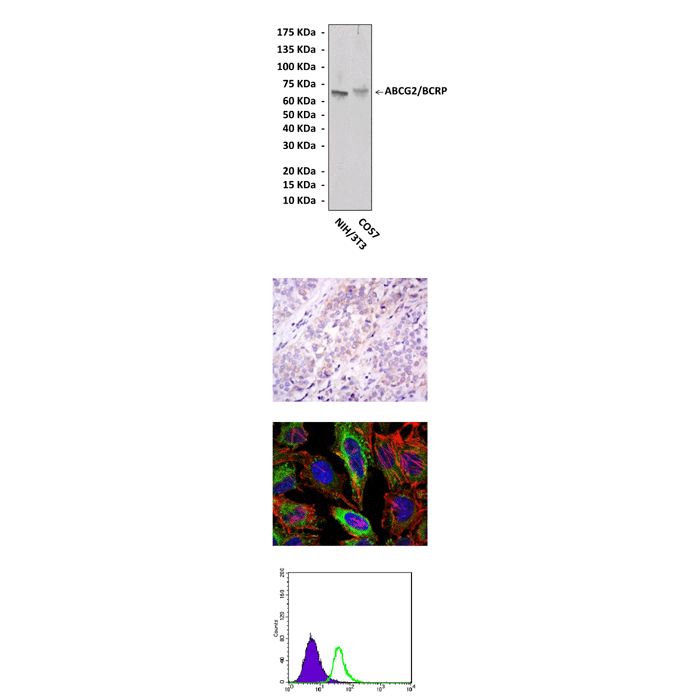
Top: Western blot detection of ABCG2 proteins in NIH3T3 and Cos7 cell lysates using ABCG2 Antibody. Middle Upper: This antibody stains paraffin-embedded human bladder cancer tissue in IHC analysis. Middle Lower: It also stains HeLa cells in confocal immunofluorescent testing (ABCG2 antibody: Green; Actin filaments: Red; DRAQ5 DNA Dye: Blue). Bottom: This antibody detects ABCG2 proteins specifically in HepG2 cells by FACS assay (ABCG2 antibody: Green; negative control: Purple).
Details
Cat.No.: | CP10386 |
Antigen: | Raised against recombinant human ABCG2 fragments expressed in E. coli. |
Isotype: | Mouse IgG1 |
Species & predicted species cross- reactivity ( ): | Human, Mouse, Monkey |
Applications & Suggested starting dilutions:* | WB 1:1000 IP n/d IHC 1:50 - 1:200 ICC 1:50 - 1:200 FACS 1:50 - 1:200 |
Predicted Molecular Weight of protein: | 70 kDa |
Specificity/Sensitivity: | Detects ABCG2 proteins in various cell lysate. |
Storage: | Store at -20°C, 4°C for frequent use. Avoid repeated freeze-thaw cycles. |
*Optimal working dilutions must be determined by end user.
Products
| Product | Size | CAT.# | Price | Quantity |
|---|---|---|---|---|
| Mouse ABCG2/BCRP Antibody: Mouse ABCG2/BCRP Antibody | Size: 100 ul | CAT.#: CP10386 | Price: $302.00 |
