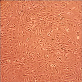
Porcine Aortic Endothelial Cells: PAOEC
Porcine Aortic Endothelial Cells (PAOEC) are primary endothelial cells isolated directly from the inner lining of the porcine aorta.
Description
Porcine Aortic Endothelial Cells (PAOEC), also referred to as porcine aorta endothelial cells (PAEC), are primary cells isolated directly from the inner lining of the porcine aorta. Unlike immortalized cell lines, these large-vessel vascular endothelial cells retain their native physiological responsive features and standard cobblestone morphology in vitro. This makes them a highly valued animal model for bridging the gap between small rodent models and human vascular biology.
Because primary cells are fastidious and sensitive to culture variations, maintaining high-quality PAOEC requires strict adherence to optimized growth conditions. These frozen cells are typically stored under liquid nitrogen to protect cell viability. Upon thawing, they are cultured using a specialized endothelial cell medium or an optimized Porcine Endothelial Cell Growth Medium formulation. Standard endothelial quality control involves morphological validation and testing for cell-specific endothelial markers, which typically includes confirming positive DiI-Ac-LDL uptake. Additionally, each lot is screened for contaminants such as mycoplasma, bacteria, yeast, and fungi.
In vivo, these cells form a continuous, single-layer antithrombotic sheet known as the endothelium, which lines the luminal surface of the porcine aorta. As the body’s largest conduit artery, the aorta subjects its endothelial lining to high hemodynamic forces, substantial hydrostatic pressure, and prominent cyclic stretch vectors generated during the cardiac cycle.
Within the multi-layered macrovascular wall architecture, these endothelial cells sit upon a thin basement membrane that separates them from the underlying tunica media, where vascular smooth muscle cells reside. While endothelial cells in smaller capillary beds experience lower pressure regimes, macrovascular PAOEC are continuously exposed to substantial shear stress. This mechanical environment strongly influences cellular orientation, causing the cells to align their long axes parallel to the direction of blood flow — a structural characteristic that can vary from the alternative alignments observed in specialized flow environments such as heart valve leaflets.
The primary function of PAOEC is to maintain vascular homeostasis, regulate endothelial homeostasis, and modulate vascular tone within the high-pressure arterial system. PAOEC synthesize and release critical vasoactive substances like nitric oxide (NO) to regulate the contraction and relaxation of adjacent smooth muscle cells. When exposed to mechanical or chemical stimuli, these cells dynamically alter the expression of NO synthase and various cellular adhesion molecules (such as VCAM-1 and ICAM-1) to regulate leukocyte trafficking and regional vascular remodeling.
Under pathological conditions, a chronic breakdown in these regulatory mechanisms leads to endothelial dysfunction, a hallmark of early-stage vascular diseases. For instance, prolonged oxidative stress — such as that induced by hydrogen peroxide () — can impair endothelial nitric oxide production and compromise overall cell viability. These primary cells respond to inflammatory and oxidative challenges by altering specific gene expression programs, which can ultimately dictate whether the endothelial monolayer maintains its barrier integrity or undergoes progressive degradation.
In biomedical and translational research, PAOEC serve as a useful model system to investigate macrovascular physiology, evaluate tissue engineering constructs, and model xenotransplantation dynamics. Because pigs share striking anatomical and physiological similarities with humans, these primary cells are widely used to study the cellular mechanics of atherosclerosis, hypertension, and fluid shear-stress kinetics. In vascular engineering, researchers frequently use co-culture configurations matching PAOEC with species-matched smooth muscle cells to study complex cellular cross-talk, which helps inform the design of implantable vascular grafts and bio-engineered heart valves.
PAOEC are utilized to explore infectious disease mechanisms and transplant biology. Investigators have used these cells to track how the replication of classical swine fever virus regulates intracellular signal transduction pathways and host gene expression. They are also central to xenotransplantation research, where studies demonstrate how direct interactions between porcine endothelial cells and human T-cells trigger the release of proinflammatory molecules during graft rejection, identifying specific cellular sensors like the tetraspanin CD82 as key drivers of xenogeneic incompatibility. By screening novel therapeutics, testing donor pretreatments (such as vasopressin), and analyzing PAOEC along with their associated products, scientists can develop targeted strategies to mitigate transplant rejection and better manage human cardiovascular disease.
- H2O2 contributes to vascular dysfunction
- Cell survival under oxidative stress depends on phosphorylation of VEGFR-3
- Replication of swine fever virus regulates signal transduction pathways and gene expression
- Direct interactions between endothelial cells and T cells trigger release of proinflammatory molecules that play a role in graft rejection
- The tetraspanin CD82 is the recognition sensor responsible for rejection of xenotransplants
- Endothelial cells from heart valves align perpendicular to flow, while aortic endothelial cells align parallel to flow, indicating the need to match the cell types when designing engineered tissue devices
- Shear stress induces changes in expression of NO synthase, VCAM-1, c-jun, MCP-1 and ICAM-1
- Swine-to-human xenotransplantation can be improved by pretreating donors with vasopressin
- Endothelial implants can be designed to increase lumen diameter and replace heart valves
Details
| Tissue | Normal healthy porcine aorta | |
|---|---|---|
| QC | No bacteria, yeast, fungi, mycoplasma | |
| Character | DiI-Ac-LDL uptake: Positive | |
| Bioassay | Attach, spread, proliferate in Growth Med | |
| Cryovial | 500,000 PAOEC (1st passage) frozen in Basal Medium w/ 10% FBS, 10% DMSO | |
| Kit | Cryovial frozen PAOEC, Growth Medium (P211-500), Subculture Rgnt Kit (090K) | |
| Proliferating | Shipped in Gr Med, 2nd psg (flasks or plates) | |
| Doublings | At least 16 | |
| Applications | Laboratory research use only (RUO). Not for human, clinical, diagnostic or veterinary use. |
Resources
FAQs
Need More Help?
Visit our comprehensive FAQ page for detailed answers to common questions.
Need More Help?
Visit our comprehensive FAQ page for detailed answers to common questions.
Primary Cell FAQs Powered by Bioz