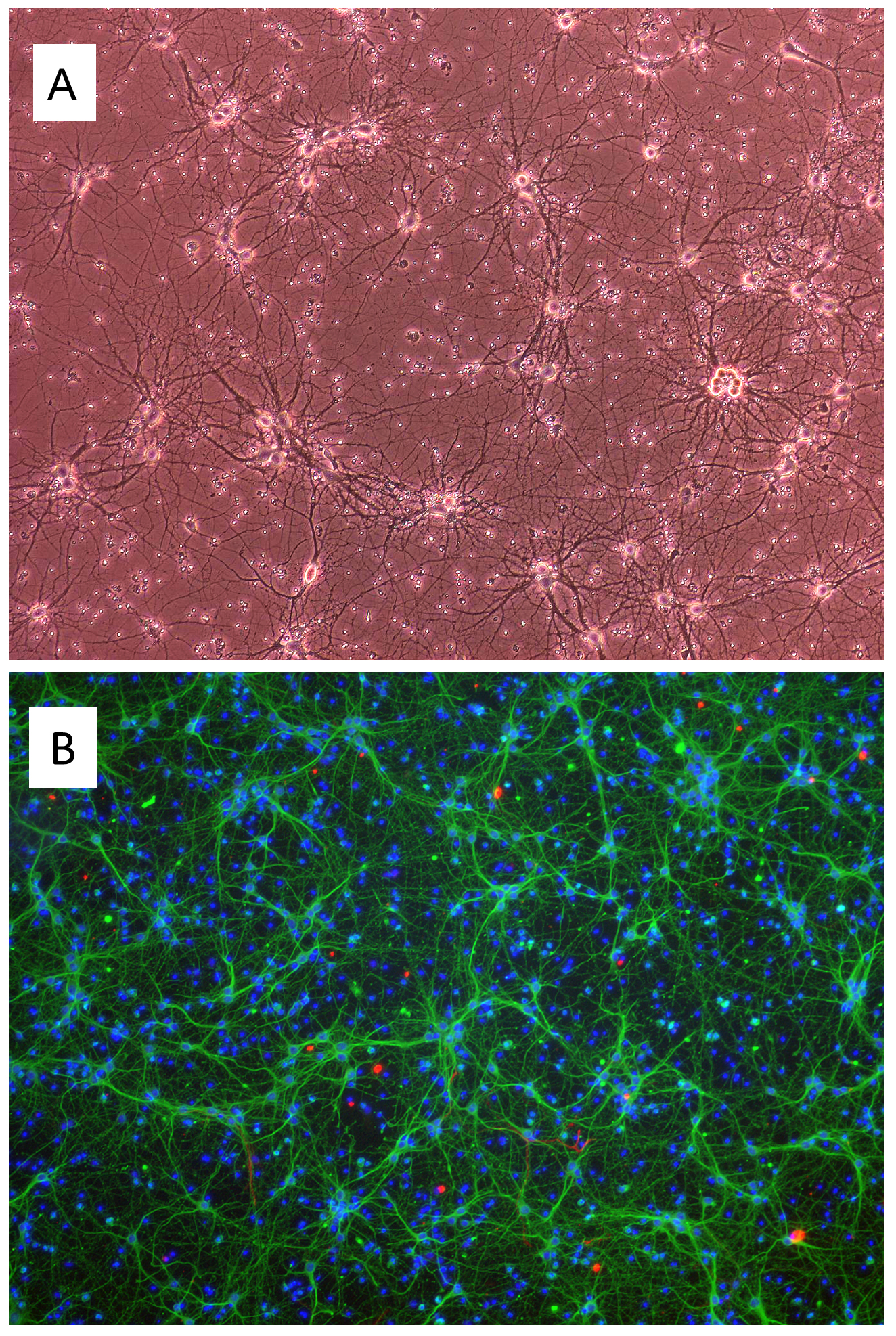
Freshly isolated Mouse Cortical Neurons (MCoN) cultured for 7 days. A: Phase contrast, 100x. B: Immunostaining for β-III Tubulin (green), GFAP (red), DAPI (blue), 100x.
Mouse Cortical Neurons: MCoN
Mouse Cortical Neurons (MCoN) are cryopreserved primary mouse neurons derived from the cerebral cortex of mouse brains.
Description
Mouse Cortical Neurons (MCoN) are cryopreserved primary mouse neurons derived from the cerebral cortex of embryonic day 18 (E18) CD1 mouse brains. As high-fidelity mouse primary cells, MCoN provide a physiologically authentic in vitro framework that mirrors native neuroanatomy far more accurately than continuous neuroblastoma or hybridoma cell lines. The cerebral cortex itself plays a key role in higher-order cognitive functions including cognition, memory, attention, perceptual awareness, thought, language, and consciousness. When cultured under optimized in vitro conditions, cells undergo rapid neurite outgrowth and network formation, expanding a complex neurite network within one week and staining positive for neuronal markers such as β‑III‑tubulin.
The structural architecture of a mature mouse cortical neuron is highly polarized, featuring a prominent cell body (soma) from which specialized processes emerge to dictate directional signaling. The dominant excitatory cell type within these isolates is the pyramidal neuron, a principal morphological class critical to the assembly of functional cortical circuits. These cortical pyramidal neurons exhibit a characteristic triangular cell body that gives rise to a single, elongated apical dendrite projecting toward upper cortical layers, several highly branched basolateral dendrites, and a single, specialized axon that propagates action potentials over long distances. In the native brain, the physiological properties, dendritic arborization patterns, and axonal projections of these neocortical pyramidal neurons vary systematically as a function of cortical depth, defining the precise laminar processing layers of the cerebral cortex. Landmark neuroanatomical studies and foundational textbooks on the synaptic organization of the brain, such as those authored by Shepherd GM, outline how these precise structural rules and spatial distributions govern the intricate input-output transformations of the mammalian neocortex.
Primary MCoN networks serve as an invaluable platform for investigating the assembly, refinement, and pathology of central nervous system pathways:
- Synaptic Transmission and Electrophysiology: Within days of seeding, developing neurons sprout neurites that seek out appropriate partners to establish the functional synapse connections required for complex neurocircuitry. This cellular layout allows investigators to dissect the precise molecular mechanics of synaptic transmission, mapping how a stimulated presynaptic neuron releases neurotransmitters across the synaptic cleft to bind postsynaptic receptors and alter the target cell’s membrane potential. By integrating advanced patch-clamp recording or multielectrode arrays (MEAs), researchers can apply targeted electrical stimulation to evaluate real-time neural firing, circuit plasticity, and how individual cells integrate a dense chorus of incoming synaptic input. The addition of specific inhibitory neurons (such as GABAergic interneurons) or glial cells into these setups allows for the reconstruction of highly balanced, homeostatic networks in vitro.
- Regional Specialization and Circuit Mapping: These primary systems are widely used to model region-specific circuitries across the cortical landscape. For example, researchers leverage MCoN to study sensory processing networks characteristic of the visual cortex, the somatosensory cortex, and the specialized barrel cortex—a region uniquely prominent in the mouse for processing tactile vibrissae (whisker) inputs. Similarly, MCoN can be configured to model the executive and motor command areas of the brain, including the frontal cortex, the prefrontal cortex, and the primary motor cortex. In descending motor pathways, the axon of a corticospinal pyramidal neuron within the motor cortex descends out of the cerebrum to interface with the spinal cord, projecting downstream to regions like the cervical spinal cord to govern precise voluntary motor control.
- Disease Modeling and High-Throughput Screening: Because cellular dysfunction and progressive degeneration within these networks are heavily implicated in various brain injuries, acute ischemic strokes, and complex neurological or psychiatric disorders, MCoN offer an excellent, non-transformed cellular model system to decode disease pathomechanisms. High-throughput platforms routinely integrate these primary cultures to perform targeted drug screening and development, developmental neurotoxicity tests, multi-color immunostaining, and long-term live cell imaging. Furthermore, by introducing fluorescent reporters or genetic vectors, researchers can monitor morphological remodeling in individually labeled neurons, tracking real-time dendritic spine remodeling and axonal degeneration under pathological stress.
MCoN (Mouse Cortical Neurons) are cryopreserved primary mouse neurons derived from the cerebral cortex of day 18 embryonic CD1 mouse brains. When cultured under the recommended conditions, MCoN arborize and form complex neurite network in one week. MCoN Stain positive for β III-Tubulin.
The cerebral cortex plays a key role in cognition, memory, attention, perceptual awareness, thought, language and consciousness. Neurons connect to other neural cells through neurites and synapses to form complex neurocircuitry for signal transmission. A healthy circuitry in the brain is essential for cognitive functions, proper control and regulation of the other parts of the body. Dysfunction and degeneration in the cerebral cortex have been implicated in various brain injuries, neurological and psychiatric disorders. Cortical neurons, therefore, provide an excellent cellular model system to study disease mechanisms and pathophysiologies. They also serve as a platform for drug screening and development, toxicity tests, immunostaining, live cell imaging, co-culturing and electrophysiology.
Details
| Tissue | Normal healthy mouse brain | |
|---|---|---|
| QC | No bacteria, yeast, fungi, mycoplasma | |
| Character | Positive for β-III Tubulin | |
| Bioassay | Plate on Poly-D-Lysine coated surface, arborize to form neurite network in Culture Med | |
| Cryovial | 2M MCoN cryopreserved after isolation in Serum-Free Freezing Medium (042-50) | |
| Kit | 2M cryopreserved MCoN, Coating Soln I (027-05), Plating Med (M817P-10), Culture Med (M817-100) | |
| Doublings | N/A – Neurons don’t proliferate in vitro | |
| Applications | Laboratory research use only (RUO). Not for human, clinical, diagnostic or veterinary use. |
Resources
FAQs
Need More Help?
Visit our comprehensive FAQ page for detailed answers to common questions.
Need More Help?
Visit our comprehensive FAQ page for detailed answers to common questions.
Primary Cell FAQs