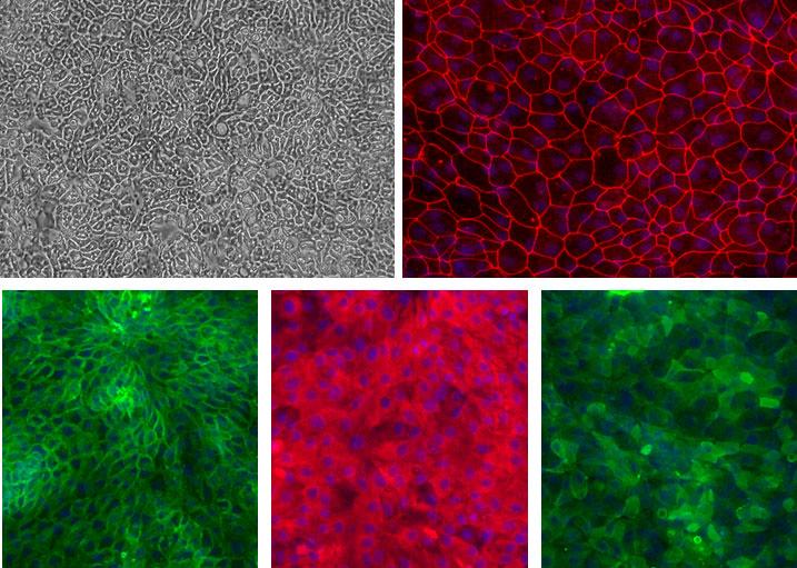
Human Duodenum Epithelial Cells (HDuEpC) and other Adult GI EpC monolayers.
Top left: Phase contrast of Ileum EpC. Top right (Red): ZO-1 of Duedenum EpC. Bottom left (Green): Na+/K+ ATPase of Colon EpC. Bottom middle (Red): pan-Cytokeratin of Gastric EpC. Bottom right (Green) Villin of Jejunum EpC. Blue: DAPI.
Human Duodenum Epithelial Cells: HDuEpC
Human Duodenum Epithelial Cells (HDuEpC) are primary isolated epithelial cells that form the continuous, microscopic lining of the human duodenum — the first section of the small intestine.
Description
Human Duodenum Epithelial Cells (HDuEpC) are primary isolated cells that form the continuous, microscopic lining of the human duodenum — the first section of the small intestine. Histologically, this specialized epithelial tissue is organized into a simple columnar epithelium, consisting of a single layer of tall, tightly packed epithelial cells designed for rapid nutrient transit and barrier integrity.
Unlike the multi-layered stratified squamous epithelium that protects areas prone to high mechanical friction (like the esophagus), this delicate epithelium is optimized for high-capacity secretion and absorption. To maintain their continuous barrier function, HDuEpCs rely on underlying active stem cells located deep within the tissue architecture to fuel rapid, ongoing cell renewal.
To understand where HDuEpCs reside, it helps to look at the broader anatomy of the proximal gastrointestinal tract. They form the cellular face of the duodenal mucosa, the specialized inner mucous membrane that directly borders the acidic chyme leaving the stomach. The tissue forms deep invaginations called the intestinal crypt (or crypts of Lieberkühn) and finger-like projections called villi. This entire tissue layer sits atop a supportive base of dense connective tissue (the lamina propria and submucosa), which houses a rich vascular network supplied by a local mesenteric artery.
Deeper still in the duodenal wall lies a layer of smooth muscle called the muscularis externa, which provides the peristaltic contractions that move digesting food. Crucially, the submucosa of the duodenum uniquely contains Brunner’s glands (brunners glands), specialized alkaline-secreting glands that protect the delicate duodenal epithelium from stomach acid.
As a primary intestinal system cell, HDuEpCs function as a dynamic, selectively permeable barrier. They allow nutrient uptake while blocking harmful luminal pathogens and toxins from breaching the body’s core. To achieve this, the stem cell population differentiates into several distinct lineages that work in tandem:
Enterocytes: The highly abundant, classic absorptive cells (or enterocytes) equipped with microscopic microvilli that maximize surface area to absorb broken-down fats, sugars, amino acids, and water.
Goblet Cell: A specialized, mucus-secreting goblet cell population that lubricates the lumen and blankets the epithelium in a thick, protective layer.
Paneth Cells: Positioned at the base of the crypts, Paneth cells secrete essential antimicrobial peptides to defend the stem cell niche.
Enteroendocrine Cells: Scattered enteroendocrine cells (or enteroendocrine cell types) act as the sensors of the gut. These specialized endocrine cell units release critical hormones like secretin and cholecystokinin (CCK) directly into the bloodstream to regulate pancreatic secretion, bile release, and gastric motility.
Tuft Cell: An chemosensory tuft cell type that acts as a sentinel to detect parasitic infections and initiate appropriate immune cascades.
M Cell: Located primarily over gut-associated lymphoid tissue, the M cell (microfold cell) samples luminal antigens and delivers them directly to underlying immune cells—such as antigen-presenting cells and T cells—to choreograph gut immunity.
Through molecular signaling between adjacent cells, this coordinated network maintains gastrointestinal homeostasis and systemic metabolic balance. Because sourcing healthy human tissue on demand is a significant logistical hurdle, isolated HDuEpCs provide a highly translatable, human-derived platform for in vitro research.
Comparative Pathophysiology
Scientists frequently utilize HDuEpCs to model acute and chronic diseases of the upper gut. They serve as a critical physiological counterpoint when compared to human gastric epithelial cell models (to study the stomach-duodenum transition) or normal colonic epithelial cells (to analyze differences along the length of the digestive tract).
Disease Modeling & Drug Screening
By culturing HDuEpCs in specialized epithelial cell growth medium, investigators can study the direct mechanisms behind cellular injury in a duodenal ulcer, nutritional malabsorption syndromes (like Celiac disease), and early-stage duodenal cancers. These primary cultures allow researchers to observe changes in cellular tight junctions and secretory pathways when exposed to inflammatory cytokines, providing an essential preclinical platform to screen novel drug compounds and therapeutic modulators before translating them to animal or clinical trials.
The intestinal epithelium is a single layer of cells organized into crypts and villi, known as the most rapidly self-renewing tissue in adult mammals. The cells that line the intestinal lumen perform the primary functions of digestion, water and nutrient absorption, and forms a barrier against luminal pathogens.
Transit-amplifying cells spend approximately two days in the intestinal crypts, dividing 4–5 times before terminally differentiating into specialized intestinal epithelial cell types. In the small intestine, the surface area is dramatically enlarged through epithelial protrusions called villi. Three days after their terminal differentiation, the cells reach the tip of the villus, undergo spontaneous apoptosis, and are shed into the gut lumen.
CAI’s intestinal epithelial culture system provides outstanding resource for investigation of intestinal epithelial cell physiology related to GI infection, inflammatory bowel disease (IBD) like Crohn’s disease, ulcerative colitis, and intestinal cancer. Our epithelial cell culture system can be efficiently used as a test platform for the potential drug candidates and disease modulators. Other applications of this culture system include functional analysis of intestinal epithelium, GI disease modeling, and regenerative therapy preclinical testing such as drug compound screening and other validation assays.
With optimized, defined culture media from CAI, the Intestinal Epithelial Cells can be seeded and maintained for as long as 8 days. Epithelial Cells grown in CAI medium form a monolayer of polarized epithelial cells with tight junction formation as evidenced by Villin (apical marker), Na+/K+ ATPase (basolateral marker), ZO-1 (tight junction marker) and pan-Cytokeratin (epithelial marker) staining.
Details
| Tissue | Normal healthy human duodenum. | |
|---|---|---|
| QC | No bacteria, yeast, fungi, mycoplasma, virus. | |
| Bioassay | Attach, spread, in Culture Medium. | |
| Cryovial | 500,000 HDuEpC in Freezing Medium. | |
| Kit | Cryovial frozen HDuEpC (732Du-05a), Culture Med (716DC-50), Coating Solution (1024-05), Thawing Solution (716T-20). | |
| Cultured | Shipped in flasks or plates in medium. | |
| Doublings | Cells do not divide and cannot be passaged. | |
| Applications | Laboratory research use only (RUO). Not for human, clinical, diagnostic or veterinary use. |
Resources
FAQs
Need More Help?
Visit our comprehensive FAQ page for detailed answers to common questions.
Need More Help?
Visit our comprehensive FAQ page for detailed answers to common questions.
Primary Cell FAQs