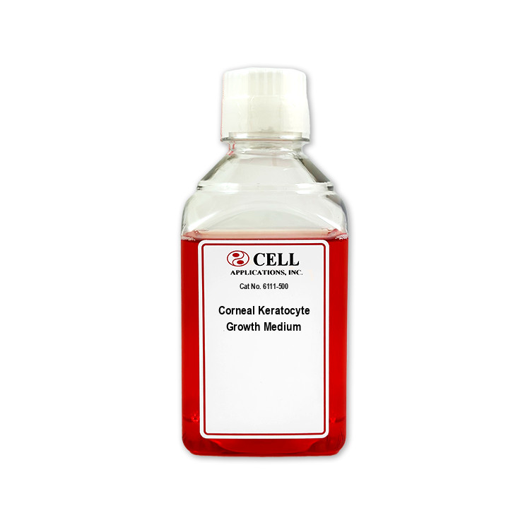
Human Corneal Keratocyte Media containing growth factors, supplements and components optimized for Human Corneal Keratocytes
Human Corneal Keratocyte Media
Corneal Keratocyte Growth Medium: All-in-one ready-to-use
Type: Growth Medium
Size: 500 ml
Price: $134.00
Description
Human Corneal Keratocyte Media are specialized cell-culture formulations designed to support the growth and maintenance of primary human corneal keratocytes (HCK), also known as corneal stromal cells. Human keratocytes are resident stromal cells located within the avascular stromal layer of the cornea, between the corneal epithelium and endothelium. They are distributed among the collagen lamellae of the stromal collagen matrix and contribute to the organization, maintenance, and repair of the corneal extracellular matrix. Through their interactions with Type I and Type V collagen, keratan sulfate proteoglycans such as keratocan, lumican, and decorin, and other matrix components, keratocytes help regulate collagen fibril diameter, fibril spacing, stromal organization, and tissue clarity. These properties are essential for corneal transparency and refractive function.
In healthy corneal tissue, keratocytes generally exhibit a quiescent phenotype and a characteristic dendritic morphology. They contribute to stromal homeostasis by maintaining the collagen matrix and expressing keratocyte-associated genes and proteins, including keratocan, lumican, and, in appropriate culture conditions, markers such as ALDH3A1. Human keratocytes also exhibit phenotypic plasticity. Following ocular trauma, wounding, infection, or corneal inflammation, they may become activated, adopt a migratory corneal-fibroblast phenotype, and, in response to signals including transforming growth factor beta (TGF-β) and platelet-derived growth factor (PDGF), differentiate further into contractile myofibroblasts. Persistent myofibroblast activation and disorganized extracellular-matrix deposition can contribute to corneal haze and loss of transparency.
Human Corneal Keratocyte Media provide a basal medium and growth supplement formulated to support the survival, attachment, expansion, and maintenance of primary HCK in vitro. The growth medium may help provide a controlled environment for studying keratocyte morphology, viability, proliferation, gene expression, extracellular-matrix production, and responses to injury or differentiation stimuli. Because keratocyte phenotype can be influenced by donor source, isolation procedure, substrate, serum or growth-factor exposure, cell density, passage number, and culture duration, the use of a specialized medium does not by itself guarantee preservation of a fully quiescent phenotype or native stromal function. These human primary-cell models support research in corneal cell biology, translational ophthalmology, wound healing, corneal inflammation, fibrosis, drug toxicity, and regenerative medicine. In ex vivo or in vitro studies, HCK may be used in:
- Monolayer & 3D Stromal Models: Single-layer cultures, three-dimensional biomimetic stromal matrices, and collagen-based tissue constructs.
- Co-Culture Systems: Multi-cell models designed to study cross-talk between keratocytes, the stratified corneal epithelium, and other ocular cell populations.
- Functional Assays: Gene-expression analysis, extracellular-matrix characterization, wound-closure/scratch assays, collagen-contraction studies, apoptosis assays, and mapping pathways that regulate myofibroblast differentiation.
Human Corneal Keratocyte Media may also be incorporated into selected models for evaluating ocular drugs, biomaterials, tissue-engineering strategies, and potential treatments for corneal scarring or haze. These models can help investigate how candidate compounds influence keratocyte activation, TGF-β- or PDGF-associated signaling, matrix remodeling, wound healing, and the transition from a quiescent keratocyte to a repair or fibrotic phenotype. They may also contribute to studies of ocular drug toxicity and inflammatory responses, although specific applications — including allergic-reaction models, stem-cell-based regenerative systems, and engineered corneal tissues — require separate validation for the complete cell culture system.
Note: These formulations are intended for research use only.
Specialized media, with ingredients and supplements optimized for the unique requirements of Human Corneal Keratocytes providing optimal culture conditions, cell health, data integrity and reproducibility.
Use with
Details
CAI media are tested for sterility in order to confirm no bacteria, yeast or fungi contaminate the solutions. The products undergo further quality control for correct pH, osmolality and lack of endotoxins. A panel of different bioassays affirm the media sustain a proper environment for expected cell-type-specific culture, growth, plating, karyotype, physiology, morphology, viability, population doublings, surface markers, cryopreservation, differentiation and/or induction.
Laboratory research use only (RUO). Not for human, clinical, diagnostic or veterinary use.
 Powered by Bioz