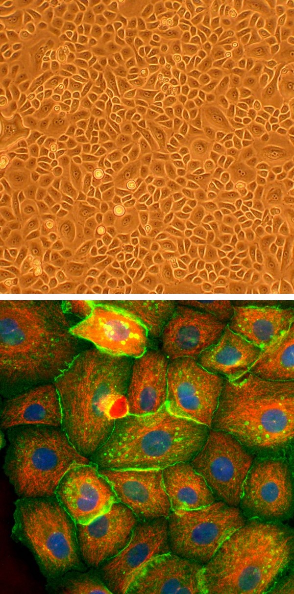
Human Corneal Epithelial Cells: HCEpC
Human Corneal Epithelial Cells (HCEpC) are highly specialized primary epithelial cells isolated from the stratified squamous epithelium that forms the outermost layer of the human cornea.
Description
Human Corneal Epithelial Cells (HCEpC) — also studied as an individual corneal epithelial cell type or as primary corneal epithelial cells — are highly specialized primary cells isolated from the stratified squamous epithelium that forms the outermost layer of the human cornea. Unlike a transformed continuous cell line, these cells of authentic human origin retain their native cell biology, functional surface receptors, and realistic physiological responsiveness ex vivo. When provided as frozen cells in a cryopreserved vial, these primary corneal cells typically exhibit high post-thaw cell viability. To maintain an optimal cell density and encourage controlled corneal epithelial proliferation within the culture vessel, investigators must utilize fully supplemented media. While epidermal growth factor is a critical mitogenic component, preventing early senescence and supporting healthy growth requires a coordinated cocktail of additional supplements.
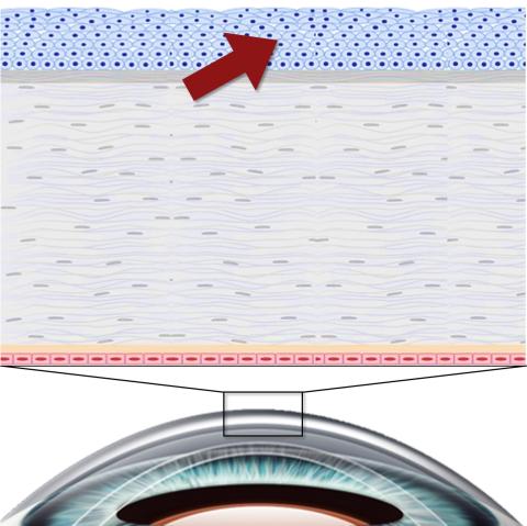
In the human body, HCEpC comprise the outermost cellular barrier of the eye, sitting directly anterior to the avascular corneal stroma and the posterior monolayer of endothelial cells. This rapidly renewing tissue maintains a smooth, transparent optical surface critical for uniform light refraction. The ongoing regenerative capacity of this layer is fueled by a specialized niche of limbal stem cells located at the limbus—the transitional border between the cornea and the sclera. As these stem cells divide, they generate transient amplifying cells that migrate centripetally and differentiate into the cuboidal basal cell layer, which subsequently matures into the superficial squamous sheets. While these corneal cells share a basic stratified barrier architecture with other mucosal systems — such as human bronchial epithelial cells and related airway epithelial cells found in the respiratory tract — HCEpC possess entirely distinct immune and structural specializations. They lack cilia and secretory goblet cells, the ciliated, mucus-secreting apparatus essential for clearing pathogens in conditions like cystic fibrosis, adapting instead for absolute optical clarity.
The primary biological function of HCEpC is to act as the front-line physical and immunological shield for the ocular surface. Through the assembly of tight junctions and desmosomal complexes, they maintain a highly selective permeability barrier that blocks the ingress of environmental toxins and infectious pathogens. They express specific tested markers of functional maturity, such as cytokeratin 3 (CK3) and cytokeratin 12 (CK12), which confirm successful cell differentiation. Furthermore, these cells maintain tissue homeostasis of cell density in vivo and coordinate rapid corneal wound healing following mechanical or chemical trauma. Upon injury, migrating cells can transiently downregulate certain differentiation markers to adopt a migratory phenotype; however, these specific marker shifts and repair behaviors are highly variable, depending heavily on the nature of the injury and localized signals from the underlying basement membrane and growth factor microenvironment.
In ophthalmic research and commercial discovery pipelines, HCEpC derived from human corneal tissue serve as a translationally rigorous model for validating topically applied therapeutics and investigating ocular surface pathologies. Safety pharmacologists utilize these cells in high-content toxicology studies to map the cytocompatibility, inflammatory liability, and structural toxicity thresholds of novel eye drops, contact lens solutions, and anti-inflammatory formulations. Numerous scientific references highlight their utility in modeling dry eye disease, mapping innate immune cascades during herpes simplex keratitis, and optimizing surgical constructs to treat limbal stem cell deficiency. By utilizing primary cultures alongside an established cell line, researchers can precisely isolate cell-type-specific variables to drive advanced regenerative medicine pipelines.
Details
| Tissue | Normal healthy human cornea | |
|---|---|---|
| QC | No bacteria, yeast, fungi, mycoplasma, virus | |
| Bioassay | Attach, spread, proliferate in Growth Med | |
| Cryovial | 500,000 HCEpC (2nd passage) frozen in Basal Medium w/ 10% FBS, 10% DMSO | |
| Kit | Cryovial frozen HCEpC, Growth Medium (211-500), Subculture Rgnt Kit (090K) | |
| Proliferating | Shipped in Tsfr Med, psg 1, flasks or plates | |
| Doublings | At least 12 | |
| Applications | Laboratory research use only (RUO). Not for human, clinical, diagnostic or veterinary use. |
Resources
FAQs
Need More Help?
Visit our comprehensive FAQ page for detailed answers to common questions.
Need More Help?
Visit our comprehensive FAQ page for detailed answers to common questions.
Primary Cell FAQs Powered by Bioz