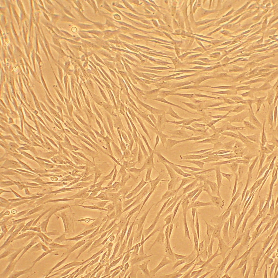
Canine Corneal Keratocytes: CnCK
Canine Corneal Keratocytes (CnCK) are specialized, neural crest-derived cells that populate the corneal stroma.
Description
Canine Corneal Keratocytes (CnCK) are specialized, neural crest-derived cells that populate the corneal stroma, playing a fundamental role in maintaining the structural integrity and transparency of the cornea. The stroma comprises the vast majority of the canine cornea’s thickness and is composed primarily of a highly organized matrix of type I collagen, alongside type V collagen which regulates fibril diameter. This structural framework is further enriched by other minor collagens (such as types III and VI) and specialized keratan sulfate proteoglycans — including keratocan, lumican, and decorin — which collectively optimize spacing to ensure minimal light scattering.
In a healthy ocular environment, these quiescent keratocytes regulate extracellular matrix (ECM) turnover. However, the tissue is frequently subject to environmental trauma and localized corneal disease, which disrupt this delicate architectural balance.
Following injury, CnCK exhibit a complex spatial and temporal response. An early wave of keratocyte apoptosis occurs immediately adjacent to the corneal lesion, mediated by epithelial-derived cytokines. Concurrently, keratocytes located more peripherally survive, proliferate, and transition into repair phenotypes (fibroblasts and myofibroblasts) to drive corneal wound healing.
These alpha-smooth muscle actin (α-SMA)-positive myofibroblasts primarily arise from resident keratocytes under the influence of transforming growth factor-beta (TGF-β) signaling following epithelial basement membrane disruption, though bone marrow-derived progenitor cells can also contribute to this pool in some inflammatory contexts. In veterinary medicine, understanding these cellular transitions is critical, as aberrant or prolonged myofibroblast activity can lead to corneal scarring, permanent corneal opacity, and visual impairment in dogs.
In the laboratory, establishing a primary cell culture of CnCK provides an invaluable in vitro model system for veterinary ophthalmology research. While continuous lines and rodent models remain widely utilized due to their high reproducibility, availability, and genetic tractability, primary canine cells offer distinct advantages for species-specific translational work.
When establishing these cultures, phenotypic stability is highly dependent on environmental conditions: standard serum-free corneal keratocyte growth media formulations help preserve the native, quiescent keratocyte state, whereas the introduction of serum shifts the cells toward fibroblastic or myofibroblastic lineages. Under optimized, phenotype-preserving conditions, primary CnCK are reported to propagate within a typical range of up to 10 population doublings, providing veterinary ophthalmologists with a high-fidelity tool to evaluate corneal patch materials, investigate tissue-specific drug toxicity, and analyze the signaling networks governing corneal regeneration.
-
Ulcerative Keratitis and Stromal Loss Pathways:
A major focus of ophthalmic research is modeling ulcerative keratitis, a highly prevalent condition in veterinary practice characterized by a deep corneal ulcer or extensive corneal ulceration. When a severe corneal lesion occurs, mechanical or infectious insults compromise the overlying corneal epithelium. This triggers an acute inflammatory cascade where bacterial proteases, host-derived matrix metalloproteinases (MMPs), and local inflammatory mediators participate in rapid stromal loss, leading to deep structural ulcers. Primary CnCK cultures allow researchers to investigate how these enzymatic pathways drive stromal melting, helping identify specific matrix inhibitors to prevent descemetocele formation or globe perforation.
-
Corneal Regeneration, Scarring, and Opacity:
During the resolution of deep corneal ulcers, the phenotype of the repair cells dictates the visual outcome. Persistent myofibroblasts generate a disorganized ECM and exert high cellular contracture, which serves as the primary driver of optical light scattering. During this transition, cells exhibit altered expression of corneal crystallins (such as aldehyde dehydrogenase); rather than directly causing the scatter, this altered crystallin profile serves as a sensitive molecular marker of phenotype change. Researchers deploy CnCK cultures to study how to modulate this response, aiming to promote true corneal regeneration over fibrotic corneal scarring to eliminate permanent corneal opacity.
-
Chronic Superficial Keratitis and Pathological Vascularization:
Healthy corneal tissue is inherently avascular to ensure transparency. In chronic inflammatory conditions, such as chronic superficial keratitis (pannus), this immune privilege is breached. Investigators use CnCK in co-culture arrays alongside vascular endothelial cells to study the mechanisms of corneal vascularization and secondary corneal pigmentation. These models reveal how activated stromal cells secrete angiogenic factors that guide the infiltration of blood vessels and melanocytes from the limbus across the cornea, providing a testing platform for novel anti-angiogenic and immunosuppressive therapies.
-
Keratoconjunctivitis Sicca and Epithelial-Stromal Crosstalk:
Proper corneal health relies on an intricate paracrine loop between the underlying stroma and the corneal epithelium, which is heavily sustained by the tear film. In diseases like keratoconjunctivitis sicca (KCS or dry eye syndrome)—including advanced neurogenic kcs where lacrimal tear glands lose autonomic innervation—the chronic lack of lubrication causes severe epithelial desiccation and secondary stromal inflammation. Researchers utilize CnCK to map how the loss of epithelial barrier protection alters stromal cell viability, drives chronic corneal degeneration, and leads to secondary ocular complications such as anterior uveitis.
-
Corneal Dystrophy, Degeneration, and Surgical Interventions:
Primary canine cultures are valuable for investigating hereditary and age-related metabolic defects, including corneal dystrophy and corneal edema. In cases of dystrophy or advanced lipid/calcium corneal degeneration, metabolic dysfunction causes abnormal opacities to accumulate within the stromal layers. When these opacities or chronic non-healing defects fail to resolve with medical therapy, a superficial keratectomy is often performed by a veterinary surgeon to physically excise the diseased tissue. Cultured CnCK serve as an empirical framework to analyze how remaining stromal layers react to surgical trauma, evaluate post-operative healing kinetics, and test the integration of bioengineered corneal matrices or mesenchymal stem cells designed to accelerate structural reconstruction.
Details
| Tissue | Normal healthy canine cornea | |
|---|---|---|
| QC | No bacteria, yeast, fungi, mycoplasma, virus | |
| Bioassay | Attach, spread, proliferate in Growth Med | |
| Cryovial | 500,000 CnCK (1st passage) frozen in Basal Medium w/ 10% FBS, 10% DMSO | |
| Kit | Cryovial frozen CnCK (Cn632-05), Growth Medium (Cn6111-500), Subculture Rgnt Kit (090K) | |
| Proliferating | Shipped in Tsfr Med, psg 1, flasks or plates | |
| Doublings | At least 15 | |
| Applications | Laboratory research use only (RUO). Not for human, clinical, diagnostic or veterinary use. |
Resources
FAQs
Need More Help?
Visit our comprehensive FAQ page for detailed answers to common questions.
Need More Help?
Visit our comprehensive FAQ page for detailed answers to common questions.
Primary Cell FAQs