


Trending Products
Products


Custom Services & Related Products
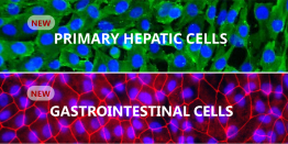
Primary Cell Services
- Human & Animal Primary Cell Isolation
- Cell-Based Assays & Custom Solutions
- 3D tissue & 3D-Bioprinting Services
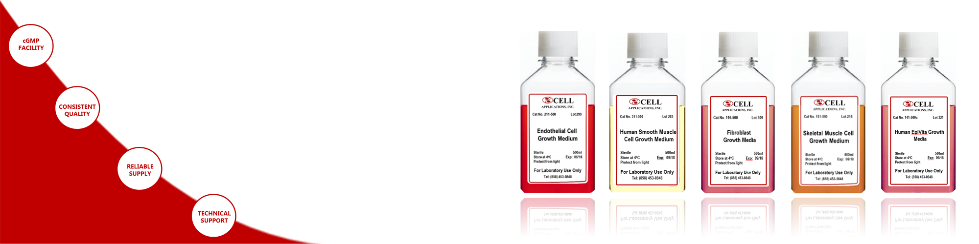
Cell Media Services
- Media Formulation & Optimization Services
- Large-scale Media Production in cGMP Facility
- Available in Standard Bottles & Flexible Bags
News & Events
Xeno-Free Media Specials
Choose Cell Applications Xeno-Free & Chemically-defined Media for your Research and Drug Discovery. Learn More
NEW! Tuft Cell Media
All-in-one ready-to-use media optimized for enrichment of Intestinal Tuft Cell precursors. Learn More
Latest Publications
Yamakami et al., 2019 J. Intern. Med
Yu et al., 2019 Mol. Cell. Endocrinology
Yang et al., 2018 Neuroscience Letters
25+
Years in Primary cell
biology business
biology business
2500+
Peer-Reviewed
Citations
Citations
40+
Countries Global
Footprint
Footprint
 Certified cgmp facility
cell culture & media
cell culture & media
Featured Customers