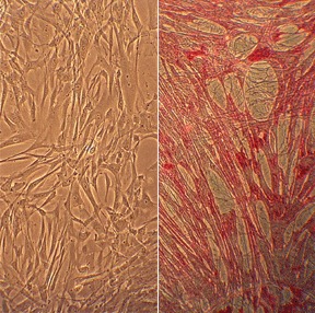
Human Umbilical Vein Smooth Muscle Cells: HUVSMC. Cells isolated from the vessel that carries oxygenated blood from the placenta to the fetus.
Human Umbilical Vein Smooth Muscle Cells: HUVSMC
Human Umbilical Vein Smooth Muscle Cells (HUVSMC) are primary smooth muscle cells isolated from the tunica media of the human umbilical vein.
Description
Human Umbilical Vein Smooth Muscle Cells (HUVSMC) are a primary cell type isolated from the tunica media of the human umbilical vein. Characterized by their expression of α-SMA alongside mature markers (such as SM22α, calponin, and smooth muscle myosin heavy chain), they provide a highly specialized in vitro system. Because α-SMA can also be expressed by myofibroblasts, confirming identity using this broader marker panel is standard practice for cultured cells. As a neonatal venous model, HUVSMC are widely utilized to study the unique physiology of the fetal-placental circuit, transcriptional regulation, and the mechanisms underlying venous remodeling.
These cells originate from the umbilical cord, which contains two umbilical arteries and one umbilical vein. In the fetal circulation, the umbilical vein serves as the single conduit carrying oxygenated, nutrient-rich blood from the placenta to the fetus.
Because HUVSMC are isolated from newborn tissue, they lack the cumulative comorbid exposures (such as aging or chronic metabolic disease) seen in adult models; however, they remain a specialized developmental model rather than a direct clinical surrogate for adult arterial disease. Because fetal vein biology—which is tuned for neonatal adaptation—does not always translate directly to adult pathophysiology, researchers often complement these human studies with animal models (such as rat or mouse venous conduits) to capture a broader range of vascular responses.
Venous smooth muscle layers are governed by ligand-receptor systems that control mural cell recruitment and structural stability. HUVSMC are central to exploring these pathways:
- Heterocellular Signaling: Remodeling is regulated by the Angiopoietin/Tie2 axis. While the Tie2 receptor is primarily localized to endothelial cells (specifically the endothelial cell layer of the tunica intima), its signaling orchestrates the recruitment and stabilization of the surrounding vascular smooth muscle cells. HUVSMC respond to these endothelial-derived cues, which regulate vascular permeability and mural cell coverage, providing a model for how these two cell types communicate to maintain venous integrity.
- Lipid-Mediated Regulation: High-density lipoproteins (HDL) are recognized for cardioprotective and anti-inflammatory properties. Studies suggest that HDL modulates signaling pathways in vascular smooth muscle cells—potentially via SR-BI interactions or through the attenuation of inflammatory transcriptional networks—to mitigate the pathological proliferation often associated with vascular disease.
HUVSMC are highly responsive and frequently utilized to investigate the pathways associated with graft failure and endovascular interventions.
- Stent and Shunt Interventions: Neointimal hyperplasia—driven by the migration and proliferation of cells—is a primary cause of shunt failure. HUVSMC are used to evaluate treatments designed to inhibit this pathological remodeling, helping researchers assess cell viability and the success of therapeutic strategies for conditions such as portal hypertension.
- Phenotypic Plasticity: A critical challenge in HUVSMC research is their tendency to dedifferentiate. Upon collagenase digestion and subsequent primary cell culture, these cells readily shift from a contractile, quiescent state toward a synthetic, proliferative phenotype. Maintaining their differentiated state requires strict control over growth factor concentrations and environmental conditions to minimize premature phenotypic drift.
In the laboratory, the maintenance of HUVSMC requires precise control over environmental factors—such as oxidative stress—to ensure consistent results. While they are a valuable tool for studying wound healing and general cell biology, they must be used with an awareness of their neonatal origins.
- Genetic Regulation: HUVSMC serve as a tool for exploring gene transcription. These vascular smooth muscle cell models have been used to demonstrate that genes coding for muscle-specific and non-muscle actin are regulated by distinct machinery, allowing cells to balance their contractile apparatus during phenotypic switching or mechanical adaptation.
- Experimental Rigor: Whether working with frozen cells or long-term cultures, investigators must carefully document passage number to avoid the impact of phenotypic dedifferentiation. By correctly utilizing these primary models and avoiding the conflation of neonatal developmental biology with adult systemic pathologies, researchers can accurately model fundamental venous interactions and test novel endovascular therapeutics.
Human Umbilical Vein Smooth Muscle Cells (HUVSMC) from Cell Applications, Inc. have been used in studies evaluating treatments that involve blood vessel stents.
- Specifically, intra-shunt delivery of motexafin gadolinium was shown to inhibit the proliferation of SMCs during treatment of portal hypertension
- Also, high-density lipoproteins (HDL) exhibit cardioprotective and anti-inflammatory properties by reducing HUVSMC expression of chemokines CCL2, CCL5, and CX3CL1, and chemokine receptors CCR2 and CX3CR1 (via NF-κB/pAkt inhibition) and by decreasing cell proliferation (via pERK inhibition), highlighting HDL importance in preventing neointimal hyperplasia, the main cause of early vein graft/stent failure
- HUVSMC were also used to demonstrate that genes coding for muscle and non-muscle actin proteins are transcribed by different transcription machinery
Characterization: positive for smooth muscle cell specific alpha-actin expression
Details
| Tissue | Normal healthy human umbilical vein | |
|---|---|---|
| QC | No bacteria, yeast, fungi, mycoplasma, virus | |
| Character | Smooth muscle specific α-actin positive | |
| Bioassay | Attach, spread, proliferate in Growth Med | |
| Cryovial | 500,000 HUVSMC (2nd passage) frozen in Basal Medium w/ 10% FBS, 10% DMSO | |
| Kit | Cryovial frozen HUVSMC (250-05n), Growth Medium (311-500), Subcltr Rgnt Kit (090K) | |
| Proliferating | Shipped in Gr Med, 3rd psg (flasks or plates) | |
| Doublings | At least 6 | |
| Applications | Laboratory research use only (RUO). Not for human, clinical, diagnostic or veterinary use. |
Resources
FAQs
Need More Help?
Visit our comprehensive FAQ page for detailed answers to common questions.
Need More Help?
Visit our comprehensive FAQ page for detailed answers to common questions.
Primary Cell FAQs Powered by Bioz