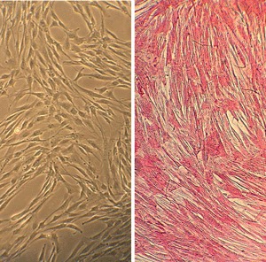
Human Subclavian Artery Smooth Muscle Cells: HScASMC. Isolated from the vessel that supplies blood to the arms.
Human Subclavian Artery Smooth Muscle Cells: HScASMC
Human Subclavian Artery Smooth Muscle Cells (HScASMC) are primary vascular cells isolated from the tunica media of the human subclavian artery.
Description
Human Subclavian Artery Smooth Muscle Cells (HScASMC) are primary vascular cells isolated from the tunica media of the human subclavian artery. Characterized by their positive expression of alpha-smooth muscle actin (α-SMA) along with a canonical panel of mature smooth muscle markers (such as SM22α, calponin, and smooth muscle myosin heavy chain), this primary cell type provides a valuable in vitro system to study macrovascular physiology.
Because they populate a high-flow conduit vessel, researchers frequently compare them to aortic smooth muscle cells to uncover regional variations along the upper arterial tree. These primary cultures serve as an anatomical model to study the molecular mechanisms driving tissue remodeling, calcification, and vascular diseases affecting the upper thoracic conduits.
In the human body, these cells reside within the muscular walls of the right and left subclavian arteries, which serve as foundational conduits for systemic blood distribution. The asymmetry of their anatomical origins is a key feature of upper thoracic architecture: the right subclavian artery branches directly from the brachiocephalic trunk (innominate artery), while the left subclavian artery arises directly from the aortic arch, positioned just distal to the left common carotid artery.
Anatomical Course and Relations
The subclavian artery travels laterally into the lower neck, arching behind the scalenus anterior (scalenus anterior muscle or anterior scalene muscle) and over the superior surface of the first rib. Classically, the vessel is divided into three distinct anatomical parts relative to its position to the anterior scalene muscle: the first part is medial to the muscle, the second part is directly deep to it, and the third part extends laterally from its lateral border.
As the third part of the subclavian artery crosses the outer border of the first rib, it becomes the axillary artery to continue its blood supply down the upper extremity. Further down the arm, the axillary artery subsequently becomes the brachial artery.
Throughout its course through the lower neck and thoracic outlet, the vessel is nested within a dense network of complex structures deep to the skin and superficial fascia:
- Nervous Structures: It passes in close proximity to the brachial plexus trunks, the phrenic nerve, the sympathetic trunk, the inferior cervical ganglion, and loops near the recurrent laryngeal nerve.
- Muscular Relations: The skeletal muscle fibers of the anterior scalene physically separate the subclavian artery from its accompanying subclavian vein.
Major Vascular Branches Before exiting the thoracic outlet, the subclavian artery gives off critical branches that supply blood to the brain, thoracic wall, and shoulder girdle:
- Vertebral Artery: Arising from the first part of the subclavian artery, it ascends through the cervical transverse foramina as a vital cervical artery, supplying the posterior circulation via the vertebrobasilar system.
- Internal Thoracic Artery: Descends along the internal thoracic wall, eventually dividing into the musculophrenic artery and superior epigastric artery.
- Thyrocervical Trunk: Gives rise to the inferior thyroid artery, the transverse scapular (suprascapular) artery, and the transverse cervical artery (from which the dorsal scapular artery highly variables and often branches, rather than arising directly from the subclavian artery itself).
- Costocervical Trunk: Typically arising from the second part of the artery, it supplies the upper intercostal spaces and deep neck muscles.
- Subscapular Artery: Arises distally off the axillary axis further down the arterial tree to supply the musculature of the scapula and chest wall.
In a healthy physiological state, this specialized vascular smooth muscle cell population maintains a highly differentiated, contractile phenotype optimized to regulate vascular tone and arterial compliance. They work in close coordination with adjacent endothelial cells—which act as the primary sensors of shear stress—to preserve endothelial integrity and maintain overall vascular and heart health.
However, in response to chronic mechanical stress, dyslipidemia, or focal endothelial injury, HScASMC exhibit significant phenotypic plasticity, transitioning to an active, synthetic state:
- Neointimal Cellularity: Under pathological stimulation, HScASMC undergo a phenotypic switch that drives hyperplasia (proliferation) and hypertrophy (increased cell size) within the arterial wall. This dysregulated proliferation contributes to increased neointimal cellularity, narrowing the blood vessel lumen during advanced plaque formation.
- Extracellular Matrix Remodeling: Upon transitioning to a synthetic phenotype, these cells increase their secretion of extracellular matrix proteins and matrix metalloproteinases (MMPs), altering the structural stability of localized obstructive lesions.
- Vascular Calcification: Under prolonged osteogenic or inflammatory stimuli, HScASMC can upregulate a bone-like gene program, accelerating mineral deposition within the extracellular matrix and reducing macrovascular elasticity.
Clinical Significance
While the subclavian artery is generally less prone to early, aggressive atherogenesis compared to the coronary artery or carotid beds, advanced systemic disease can cause severe stenosis. Subclavian artery stenosis can restrict downstream flow to the arm or induce subclavian steal syndrome, where retrograde flow is pulled down from the vertebral artery to supply the upper limb, potentially compromising vertebrobasilar perfusion. Because sourcing fresh, healthy human arterial tissue is clinically restricted, isolated HScASMC provide an invaluable human-derived platform to evaluate how smooth muscle cells from the upper arterial tree respond to therapeutic interventions.
Researchers leverage this specific cell type in vitro to test anti-proliferative drugs, vasoactive agents, or novel endovascular stent coatings designed to suppress hyperplastic occlusion. By optimizing culture parameters, such as the exact composition of the smooth muscle cell medium or controlling the initial plating density, investigators can accurately simulate macrovascular disease states without confounding variables, accelerating the development of targeted therapies for peripheral artery disease.
Human Subclavian Artery Smooth Muscle Cells (HScASMC) provide a useful in vitro system to study all aspects of cardiovascular function and disease, especially those related to mechanisms of hyperplasia and hypertrophy of intimal smooth muscle cells leading to vascular occlusion.
Characterization: Positive for smooth muscle cell specific alpha-actin expression
Details
| Tissue | Normal healthy human subclavian artery | |
|---|---|---|
| QC | No bacteria, yeast, fungi, mycoplasma, virus | |
| Character | Smooth muscle specific α-actin positive | |
| Bioassay | Attach, spread, proliferate in Growth Med | |
| Cryopvial | 500,000 HScASMC (2nd passage) frozen in Basal Medium w/ 10% FBS, 10% DMSO | |
| Kit | Cryovial frozen HScASMC (3510-05a), Grwth Med (311-500), Subcltr Rgnt Kit (090K) | |
| Proliferating | Shipped in Gr Med, 3rd psg (flasks or plates) | |
| Doublings | At least 12 | |
| Applications | Laboratory research use only (RUO). Not for human, clinical, diagnostic or veterinary use. |
Resources
FAQs
Need More Help?
Visit our comprehensive FAQ page for detailed answers to common questions.
Need More Help?
Visit our comprehensive FAQ page for detailed answers to common questions.
Primary Cell FAQs