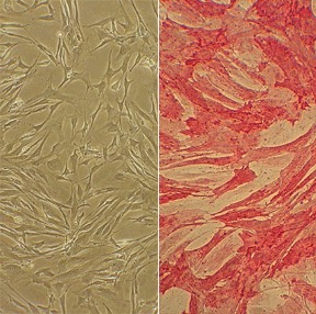
Human Pulmonary Artery Smooth Muscle Cells: HPASMC. Cells isolated from the vessel that carries deoxygenated blood from the heart to the lungs.
Human Pulmonary Artery Smooth Muscle Cells: HPASMC
Human Pulmonary Artery Smooth Muscle Cells (HPASMC) are primary smooth muscle cells isolated from the tunica media of human pulmonary arteries.
Description
Human Pulmonary Artery Smooth Muscle Cells (HPASMC) are primary smooth muscle cells isolated from the tunica media of human pulmonary arteries. Characterized by their positive expression of α smooth muscle actin (α-SMA), HPASMC serve as an essential in vitro model system.
To ensure high culture purity, these cells are checked against a panel of tested markers to confirm they are free of contamination. In research, HPASMC allow investigators to study the unique physiology and pathophysiology of the pulmonary vasculature, exploring the mechanisms that drive proliferation, migration, and hyper-reactivity.
In vivo, these cells populate the walls of the pulmonary artery and its downstream branches, forming a low-pressure, high-compliance vascular bed. This system is responsible for delivering deoxygenated blood from the right ventricle to the lungs for oxygenation.
Unlike systemic arteries, the pulmonary vasculature exhibits a distinct response to alveolar hypoxia. When oxygen levels drop in the lungs, it triggers hypoxic pulmonary vasoconstriction to redirect blood to better-ventilated areas. However, sustained or chronic lung hypoxia leads to persistent vasoconstriction and structural remodeling of the vessel walls, driving phenotypic switching in PASMC.
This specialized vascular niche separates pulmonary pathology from other chronic respiratory diseases like asthma. While asthma primarily remodels the airways, chronic hypoxic stress specifically alters the pulmonary vascular bed, driving pulmonary vascular disease and transforming the underlying vascular smc populations.
The primary function of HPASMC is to regulate pulmonary vascular tone and manage perfusion. Under pathological conditions, however, these cells undergo a profound phenotypic switch that drives pulmonary arterial hypertension (PAH)—including its severe, non-hereditary form, idiopathic pulmonary arterial hypertension (IPAH).
During PAH progression, pulmonary arterial walls thicken because of smooth muscle hypertrophy and abnormal pasmc proliferation. This hyperproliferative, apoptosis-resistant state narrows the vessel lumen, increases pulmonary vascular resistance, and can ultimately lead to right ventricular failure.
Researchers utilize HPASMC to study the specific signaling pathway networks that orchestrate this smooth muscle cell proliferation and pasmc hyperplasia. Key areas of investigation include:
- Mitogenic & Growth Factor Cascades: Chronic exposure to canonical growth factor molecules (such as PDGF and TGF-β), endothelin-1, and serotonin stimulates HPASMC growth. Under conditions of low oxygen (hypoxia), these proliferative pathways are significantly amplified compared to baseline oxygen levels (normoxia).
- Genetic Modifications: Mutations in bone morphogenetic protein receptor type 2 (BMPR2) alter downstream protein expression profiles, disrupting normal anti-proliferative signaling and lowering the threshold for hyper-proliferation. While they exhibit altered differentiation potential, they remain classified as vascular lineages distinct from multipotent stem cells.
- Inflammatory Cross-Talk: Circulating cytokines like IL-6 alter the transcriptional landscape of HPASMC, promoting survival and extracellular matrix deposition. Other exploratory factors, such as Interleukin-22 (IL-22), are also being investigated for their potential to promote cell growth via NADPH oxidase-dependent pathways.
HPASMC are a cornerstone of preclinical drug discovery for halting the progression of devastating vascular diseases.
In Vitro Modeling & Assay Optimization
In the laboratory, HPASMC are maintained in a specialized, commercially optimized smooth muscle cell medium. Because primary cell behavior can drift over time, researchers rely on advanced formulations of growth media and tightly control parameters like plating density to keep the cells in a stable, differentiated state during experimental passaging.
Therapeutic Screening
By comparing how cells from healthy donors and patients with IPAH behave in vitro, scientists can test targeted small molecules and gene-regulation strategies. These models are heavily leveraged to identify selective, antiproliferative compounds capable of suppressing vascular disease and reducing thickened arterial walls, while sparing normal systemic vessels.
Human Pulmonary Artery Smooth Muscle Cells (HPASMC) from Cell Applications, Inc. have been used to show that:
- Antitumor drugs can selectively target remodeled pulmonary vessels, but not normal vessels
- Serotonin induces their growth and activates the BMP receptor
- IL-22 promotes the growth of pulmonary vascular SMCs via a signaling mechanism that involves NADPH oxidase-dependent oxidation
- Inducers of pulmonary hypertension, characterized by thickened pulmonary arterial walls, can be suppressed by targeting gene transcription
- Cell growth can be suppressed by trans-retinoic acid by inducing expression of a known cell growth suppressor,
- Retinoic acid and its receptors may be involved in cell growth and pulmonary vascular remodeling
- Iron chelation exerts effects on vascular remodeling and is implicated for development of pulmonary hypertension as a result of ROS activity
Characterization: positive for smooth muscle cell specific alpha-actin expression
Details
| Tissue | Normal healthy human pulmonary artery | |
|---|---|---|
| QC | No bacteria, yeast, fungi, mycoplasma, virus | |
| Character | Smooth muscle specific α-actin positive | |
| Bioassay | Attach, spread, proliferate in Growth Med | |
| Cryovial | 500,000 HPASMC frozen in Basal Medium w/ 10% FBS, 10% DMSO | |
| Kit | Cryovial frozen HPASMC (352-05a), Growth Medium (311-500), Subcltr Rgnt Kit (090K) | |
| Proliferating | Shipped in Gr Med, 3rd psg (flasks or plates) | |
| Doublings | At least 16 | |
| Applications | Laboratory research use only (RUO). Not for human, clinical, diagnostic or veterinary use. |
Resources
FAQs
Need More Help?
Visit our comprehensive FAQ page for detailed answers to common questions.
Need More Help?
Visit our comprehensive FAQ page for detailed answers to common questions.
Primary Cell FAQs Powered by Bioz