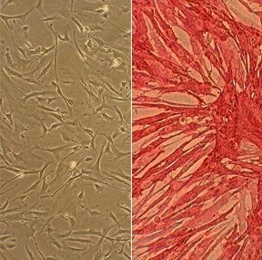
Human Internal Thoracic / Mammary Artery Smooth Muscle Cells: HITASMC. Cells isolated from the vessel that supplies the anterior chest wall and breasts.
Human Internal Thoracic Artery Smooth Muscle Cells: HITASMC
Human Internal Thoracic Smooth Muscle Cells (HITASMC) are primary human smooth muscle cells isolated from the tunica media of healthy internal thoracic arteries.
Description
Human Internal Thoracic Smooth Muscle Cells (HITASMC), also called internal mammary artery smooth muscle cells, are primary human smooth muscle cells isolated from the tunica media of healthy internal thoracic arteries. These human primary cells robustly express contractile markers such as α smooth muscle actin (α-SMA), SM22α, and calponin.
In cardiovascular research, HITASMC serve as a baseline model for studying macrovascular smooth muscle biology and arterial resistance to atherosclerosis and remodeling. They are widely utilized in vitro to investigate how an artery’s specific genetic and phenotypic profile correlates with its relative resistance to accelerated arterial disease and pathological vascular remodeling.
In vivo, these vascular smooth muscle cells are located within the internal thoracic artery (ITA). The internal thoracic artery arises from the subclavian artery and descends along the inner anterior thoracic wall, deep to the costal cartilages and internal intercostal muscles. It gives rise to pericardiacophrenic artery, anterior intercostal, and perforating branches, and typically ends as the musculophrenic artery (musculophrenic atery) and superior epigastric artery. The internal thoracic artery resides within a vascular bed that is relatively resistant to atherosclerosis. It is well suited to arterial flow and is lined by a highly functional endothelial cell layer that works in tandem with the underlying vascular smc population to maintain structural integrity. This protective vascular microenvironment makes the ITA more resistant to atherosclerosis than many other arterial beds, such as the coronary artery or branches of a posterior intercostal artery, which are far more prone to chronic plaque development. The primary function of HITASMC is to regulate vascular tone and maintain arterial wall elasticity. However, their clinical fame stems from their performance in surgical conduits. The internal thoracic artery is relatively resistant to atherosclerosis and is the preferred arterial conduit for CABG. When utilized as ITA grafts, these vessels display superior long-term patency rates. Unlike other conduits, they exhibit a striking resistance to intimal thickening and downstream vascular occlusion after implantation. In vitro, HITASMC often show lower proliferative and migratory responses to mitogens such as PDGF-BB than smooth muscle cells from more disease-prone vessels, making them useful for studying mechanisms of vascular resilience. Researchers use this passage of cells to study how they regulate intracellular networks—such as the localized coordination of store-operated calcium entry (SOCE) and phosphodiesterases (like PDE1C) at the cell’s leading edge—to prevent the uncontrolled migration that typically drives restenosis in other vessels.
Because sourcing fresh, healthy human arterial tissue is clinically restricted, isolated HITASMC provide an invaluable human-derived in vitro platform to study the molecular mechanisms of vascular health.
Comparative Vascular Biology
Investigators frequently utilize HITASMC alongside cells from disease-prone vascular beds (such as coronary artery smooth muscle cells) or models of vascularpulmonary biology (such as the human pulmonary artery, often studied in the context of pulmonary hypertension) to map the tissue-specific factors that dictate vascular resilience. These comparisons help researchers isolate whether protective vascular traits are driven by intrinsic differences in the smooth muscle cell type or by specialized cross-talk with adjacent endothelial cells.
In Vitro Assays and Quality Control
In the laboratory, HITASMC are maintained in specialized smooth muscle cell medium optimized to preserve their differentiated phenotype. These cultures are highly valuable for testing targeted therapeutics; for instance, researchers can introduce a neutralizing human IL-6 or IL-6 receptor antibody into the culture system as an in vitro tool to block specific inflammatory signaling pathways and observe downstream changes in cell behavior. To ensure experimental validity, researchers strictly screen these primary lines for common cellular contaminants, such as mycoplasma, protecting the integrity of long-term signaling and genomic profiling assays.
Human Internal Thoracic Smooth Muscle Cells (HITASMC) are derived from the normal internal thoracic arteries (formerly called internal mammary arteries).
HITASMC, along with Human Coronary Artery Smooth Muscle Cells, both from Cell Applications, Inc. have been used to investigate the gene expression differences between smooth muscle cells from different arteries, underlying their differential response to injuries and proliferation stimuli.
Characterization: positive for smooth muscle cell specific alpha-actin expression
Details
| Tissue | Normal healthy human intrathoratic artery | |
|---|---|---|
| QC | No bacteria, yeast, fungi, mycoplasma, virus | |
| Character | Smooth muscle specific α-actin positive | |
| Bioassay | Attach, spread, proliferate in Growth Med | |
| Cryovial | 500,000 HITASMC (2nd passage) frozen in Basal Medium w/ 10% FBS, 10% DMSO | |
| Kit | Cryovial frozen HITASMC (358-05a), Growth Medium, (311-500), Subcltr Rgnt Kit (090K) | |
| Proliferating | Shipped in Gr Med, 3rd psg (flasks or plates) | |
| Doublings | At least 8 | |
| Applications | Laboratory research use only (RUO). Not for human, clinical, diagnostic or veterinary use. |
Resources
FAQs
Need More Help?
Visit our comprehensive FAQ page for detailed answers to common questions.
Need More Help?
Visit our comprehensive FAQ page for detailed answers to common questions.
Primary Cell FAQs Powered by Bioz