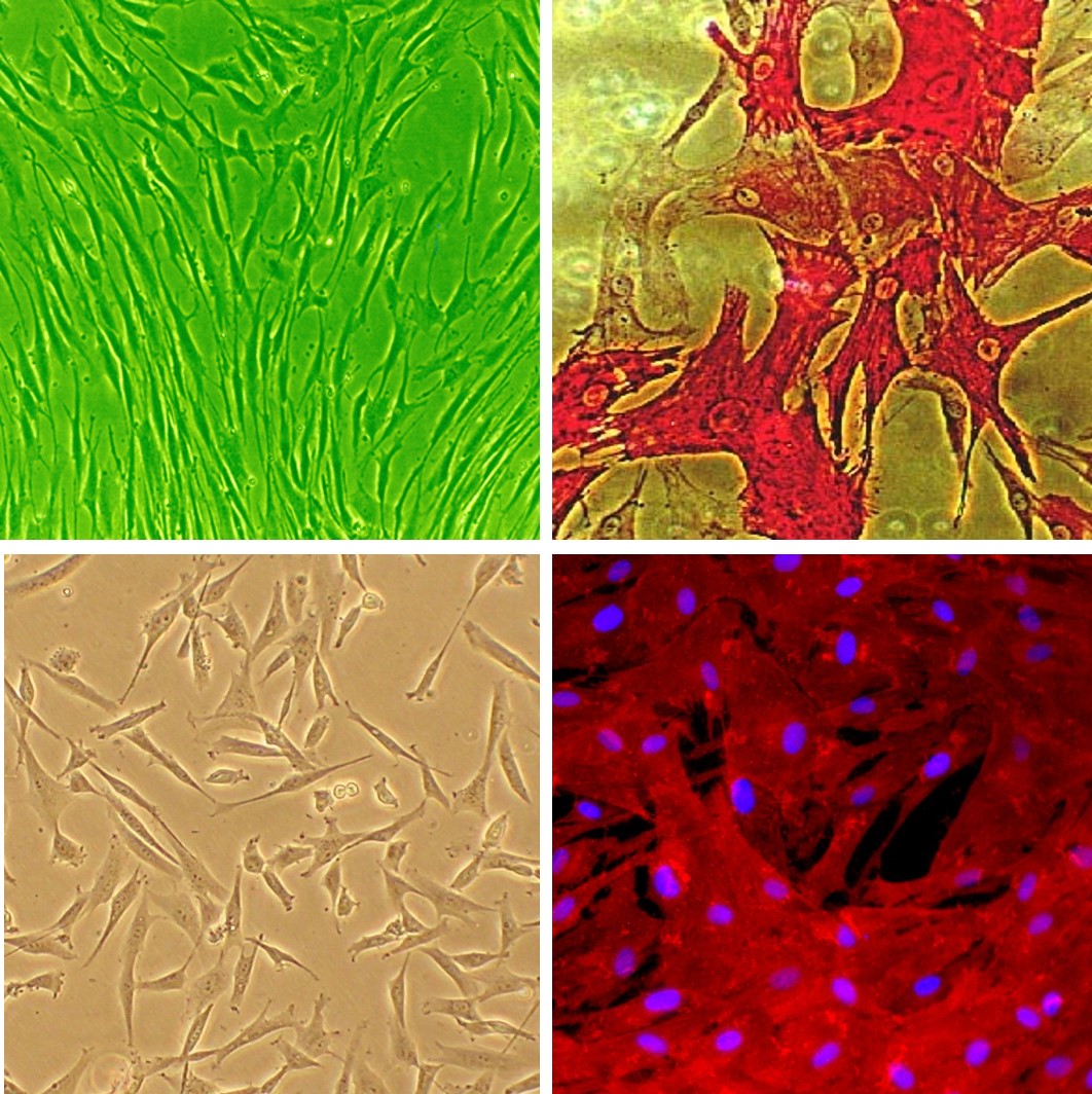
Human Coronary Artery Smooth Muscle Cells: HCASMC
Human Coronary Artery Smooth Muscle Cells (HCASMC) are specialized vascular smooth muscle cells (VSMCs) isolated from the tunica media of healthy human coronary arteries.
Description
Human Coronary Artery Smooth Muscle Cells (HCASMC) are specialized vascular smooth muscle cells (VSMCs) isolated from the tunica media of healthy human coronary arteries. Within the coronary vasculature, these cells represent a critical cell type responsible for maintaining structural integrity, regulating local vascular tone, and modulating myocardial blood flow through coordinated contraction and relaxation. Characterized by an elongated, “hill-and-valley” multilayered pattern at post-confluence in vitro — a morphological feature characteristic of many cultured vascular smooth muscle sources — HCASMC exhibit remarkable phenotypic plasticity.
Unlike transformed continuous cell lines, primary HCASMC natively maintain a panel of mature contractile markers, including smooth muscle myosin heavy chain (MYH11), smoothelin, calponin, SM22α (TAGLN), and alpha-smooth muscle actin (-SMA). However, their phenotype is highly sensitive and dynamically influenced by substrate stiffness, cyclic stretch, and the local biochemical milieu. In response to mechanical trauma, hypoxia, or environmental cues, they can transition from a quiescent, contractile state to an active, synthetic, and migratory phenotype, downregulating these contractile proteins while upregulating matrix synthesis. To sustain their native properties and avoid premature senescence in vitro, they require optimized smooth muscle growth media and specialized attachment factors, and they are frequently evaluated alongside appropriate non-vascular negative control cell types, such as dermal or tissue-matched fibroblasts.
In the living organism, these cells are arranged circumferentially in concentric layers within the tunica media, sandwiched between the inner endothelial cell monolayer (the tunica intima) and the outer adventitial connective tissue of the coronary arteries. Developmentally, coronary VSMCs derive from multiple embryologic sources depending on the vascular bed and species — including epicardial-derived cells and neural crest derivatives — rather than a single, uniform progenitor population. Sourced directly from the human coronary bed — where progressive vessel obstruction directly precipitates myocardial ischemia and ischemic heart disease — primary HCASMC provide a translationally rigorous human platform. They are frequently compared to distinct vascular beds, such as cardiac microvascular endothelial cells or internal thoracic artery smooth muscle cells, to map structural and functional heterogeneity. Because their performance is highly donor-dependent, utilizing authenticated and validated fibroblasts or healthy smooth muscle donor lots free from mycoplasma is critical for experimental precision.
The primary biological function of HCASMC in vivo is to maintain structural compliance and dynamically regulate blood pressure through vasoconstriction and vasodilation. However, under pathological conditions, their phenotypic switching plays a dual role in cardiovascular disease. In the microenvironment of a developing plaque, synthetic HCASMC migrate into the tunica intima, proliferating to form a protective fibrous cap over atherosclerotic plaques to stabilize the lipid core. Conversely, chronic inflammatory signaling can drive these smooth muscle cells to undergo osteogenic transdifferentiation—a pathological shift where they downregulate contractile proteins, alter their gene expression profiles, and actively deposit calcium and phosphate into the vessel wall, culminating in severe vascular calcification and arterial stiffening.
In cardiology, discovery pharmacology, and tissue engineering, HCASMC function as a premier human-background platform. Investigators widely deploy these primary cultures to dissect the molecular networks driving coronary artery disease (CAD). For instance, researchers use them to model ischemia-reperfusion injuries, map downstream cytokine networks, or test targeted therapeutics using species-matched human interleukin-6 neutralizing antibodies or cross-reactive blocking reagents. To map potential macrovascular toxicity and ensure target selectivity, candidate small molecules are often evaluated across a comprehensive vascular screening panel that includes HCASMC, appropriate endothelial models, and cardiac myocytes. When evaluating complex vascular responses in co-culture systems pairing HCASMC with endothelial cell lines or primary endothelial cells, investigators utilize meticulously optimized or compartmentalized media combinations that include human coronary artery smooth muscle cell media and endothelial cell growth media to support both cell types without inducing phenotypic loss.
Furthermore, because coronary occlusions often require structural stenting or surgical bypass, bioengineers leverage HCASMC to design advanced biomaterial interfaces, test stem cell differentiation kits for vascular graft endothelialization, and evaluate drug-eluting stent (DES) coatings. By tracking how primary smooth muscle cells respond to anti-proliferative agents, researchers can precisely design interventional therapies that suppress hyperplastic smooth muscle expansion within the injured vessel lumen without stalling critical endothelial layer recovery.
- Study signaling pathways regulating smooth muscle differentiation and chronic inflammation of arterial wall that leads to artherosclerosis
- Demonstrate that STAT-1 and STAT-3 regulate VEGF production in smooth muscle cells by having opposing effects on HIF-1α expression
- Examine the mechanisms of hypoxia and reoxigenation injuries in by demonstrating increased production of ROS and inflammatory cytokines, and further showing that DHA is not beneficial in this type of injuries
- Investigate (by also using human Internal Thoracic Artery Smooth Muscle Cells obtained from Cell Applications, Inc.), the gene expression differences between smooth muscle cells from different arteries, underlying their differential response to injuries and proliferation stimuli
- Suggest the hypermethylation of SOCS3 gene as the connection between TNF-α and IGF-1 released in response to mechanical injury during coronary intervention, and the induction of cytokines leading to intimal hyperplasia and restenosis
- Develop a novel VEGFR/MET-targeted inhibitor with improved antitumor efficacy and decreased toxicity
- Investigate novel therapies and drug combinations to achieve optimal target selectivity
- Generate elastic scaffolds for tissue engineering and novel treatment strategies to prevent stent restenosis by designing new materials, or drug therapies to preferentially inhibit smooth muscle cell growth
Details
| Tissue | Normal healthy human coronary artery | |
|---|---|---|
| QC | No bacteria, yeast, fungi, mycoplasma, virus | |
| Character | Smooth muscle specific α-actin positive | |
| Bioassay | Attach, spread, proliferate in Growth Med | |
| Cryovial | 500,000 HCASMC (2nd passage) frozen in Basal Medium w/ 10% FBS, 10% DMSO | |
| Kit | Cryovial frozen HCASMC (350-05a), Growth Medium (311-500), Subcltr Rgnt Kit (090K) | |
| Proliferating | Shipped in Gr Med, 3rd psg (flasks or plates) | |
| Doublings | At least 16 | |
| Applications | Laboratory research use only (RUO). Not for human, clinical, diagnostic or veterinary use. |
Resources
FAQs
Need More Help?
Visit our comprehensive FAQ page for detailed answers to common questions.
Need More Help?
Visit our comprehensive FAQ page for detailed answers to common questions.
Primary Cell FAQs Powered by Bioz