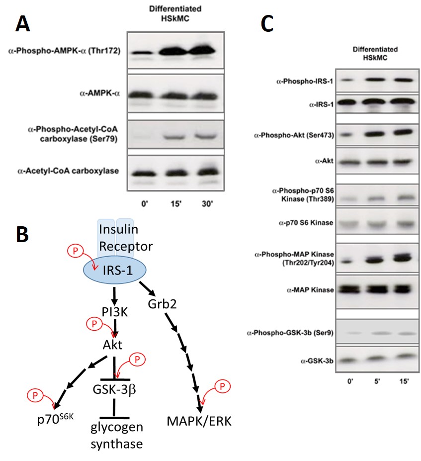
Human Skeletal Muscle Cells (HSkMC) response to forced activation of AMPK, leading to ACC phosphorylation. Cells serum-starved 16 hr & treated for 0, 15 or 30 min w/ 100nM oligomycin, which inhibits oxidative phosphorylation & causes activation of AMPK (A). Simplified insulin signal transduction map. Specific phosphorylation events examined in Fig. C are marked
...Human Skeletal Muscle Cells: S-HSkMC: Pre-Screened
Human Skeletal Muscle Cells (S-HSkMC) are primary cells isolated from human skeletal muscle tissue pre-screened for Insulin & AMPK signaling.
Description
Human Skeletal Muscle Cells (S-HSkMC) — alternatively referred to as primary human myoblasts, skeletal muscle progenitor cells, or satellite cell-derived myoblasts — are primary cells isolated from human skeletal muscle tissue. While biopsies from skeletal muscle groups such as the quadriceps, vastus lateralis, or pectoral girdle serve as common tissue sources, these mononucleated cells exist as proliferative, mononuclear myoblasts in their undifferentiated state. Unlike an immortalized cell line, primary S-HSkMC provide a translationally rigorous human model. To address donor-to-donor variability, specific lots undergo quality control to verify they retain the key features of human muscle tissue, including the ability to undergo uniform skeletal muscle differentiation into multinucleated myotubes and exhibit robust signaling in metabolic assays. For optimal results, they require a specialized skeletal muscle cell medium to support healthy cell culture, preventing confusion with unrelated environments like smooth muscle cell medium.
The differentiation of S-HSkMC follows a tightly choreographed program that transforms individual myogenic cells into functional, multinucleated contractile structures.
- Syncytium Formation: Upon mitogen withdrawal, mononuclear myoblasts align, elongate, and fuse into mature muscle fibers. This process recapitulates native mechanisms of muscle differentiation, skeletal muscle regeneration, and overall skeletal muscle growth cell kinetics.
- Sarcomeric Assembly: Structural maturation is marked by the appearance of organized contractile machinery, including parallel arrays of actin and myosin. This shift is coordinated by a precise wave of gene expression involving factors like MyoD and myogenin, alongside structural components such as dystrophin—a protein critically disrupted in muscular dystrophy. Cell surface components, such as a heparan sulfate proteoglycan matrix, help stabilize these assemblies, which can be visualized via immunofluorescence microscopy.
As skeletal muscle cells are the primary quantitative tissue driver of postprandial glucose disposal, maintaining functional signaling cascades is vital for metabolic research.
- The AMPK Sensor: These skeletal muscle cells are pre-screened to ensure a functional AMP-activated protein kinase (AMPK) cascade. Acting as an energy sensor, AMPK mimics the metabolic stress of contraction by phosphorylating downstream targets like acetyl-CoA carboxylase (ACC), managing the balance between lipid storage and oxidation.
- Insulin Cascade Fidelity: Upon insulin binding, the insulin receptor autophosphorylates, recruiting insulin receptor substrate-1 (IRS-1) to propagate the signal through the PI3K/Akt pathway. This cascade orchestrates the translocation of GLUT4 vesicles to the plasma membrane, facilitating glucose uptake. The verified presence of this uncompromised cascade is essential for investigators seeking to model metabolic dysfunction.
The robust handling profile of this cell type enables reproducible discovery workflows across advanced biomedical and therapeutic pipelines.
- Disease Modeling: Researchers utilize these muscle cells in cell culture to model type 2 diabetes and cellular insulin resistance, inducing defects via chronic exposure to free fatty acids or pro-inflammatory cytokines.
- Regeneration and Atrophy: These systems serve as a human platform for investigating muscle-wasting mechanisms (sarcopenia and cachexia). Pharmacologists challenge myotubes with catabolic stimuli to screen therapeutic candidates designed to preserve mass, activate native skeletal muscle satellite cells (muscle stem cells) to enhance muscle regeneration, and activate downstream pathways. This specialized somatic profile sets them apart from smooth muscle cells, which govern contraction in vascular networks or hollow organs (e.g., the human esophagus).
Human Skeletal Muscle Cells: S-HSkMC Pre-Screened for Insulin & AMPK signaling are isolated from the skeletal muscle of hamstrings and retain morphological, biochemical, and metabolic characteristics of skeletal muscle. Pre-screened HSkMCs can undergo differentiation to exhibit actin and myosin myofilaments and are specially tested for functional AMPK & Insulin Signaling Pathways.
AMPK Signaling Pathway
AMP-activated protein kinase (AMPK) is a major cellular regulator of lipid and glucose metabolism and mediates the metabolic changes associated with exercise. AMPK phosphorylates and inhibits activity of acetyl CoA carboxylase (ACC), the enzyme responsible for making malonyl-CoA which is required for fatty acid chain elongation.
Insulin Signaling Pathway
Skeletal muscle is one of major target tissue of insulin action. Upon insulin binding, insulin receptor tyrosine kinases catalyze autophosphorylation of tyrosine residues providing docking sites for downstream signaling components, such as IRS-1 and Grb2, which relay the signaling further into the cell (see Fig.B).
Details
| Tissue | Normal healthy human limb skeletal muscle | |
|---|---|---|
| QC | No bacteria, yeast, fungi, mycoplasma, virus | |
| Cryovial | 500,000 HSkMC (2nd passage) frozen in Basal Medium w/ 10% FBS, 10% DMSO | |
| Kit | Cryovial frozen HSkMC (150-05f), Growth Medium (151-500), Subcltr Rgnt Kit (090K) | |
| Proliferating | Shipped in Gr Med, 3rd psg (flasks or plates) | |
| Doublings | At least 15 | |
| Applications | Laboratory research use only (RUO). Not for human, clinical, diagnostic or veterinary use. |
Resources
FAQs
Need More Help?
Visit our comprehensive FAQ page for detailed answers to common questions.
Need More Help?
Visit our comprehensive FAQ page for detailed answers to common questions.
Primary Cell FAQs Powered by Bioz